A turning point for young neurons

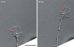
During neural development, newborn neurons extend axons toward distant targets then form connections with other cells. This process depends on the growth cone, a dynamic structure at the growing axon tip of the neuron that detects attractive and repulsive guidance cues. Many axon guidance molecules have been identified, and their functions are well characterized, but exactly how they cause growth cone turning has been unclear.
Hiroyuki Kamiguchi of the RIKEN Brain Science Institute, Wako, and his colleagues have now shown that repulsive growth cone turning is driven by a process called endocytosis1, whereby portions of the growth cone’s membrane are removed and internalized.
Endocytosis occurs continuously in all neurons to remove receptors and other membrane proteins for recycling, and to take-up neurotransmitters after their release. It is mediated by a molecule called clathrin, which induces formation of spherical ‘pits’ in the membrane that are then pulled into the cell.
By tagging clathrin with a fluorescent marker and observing the cells under a microscope, Kamiguchi’s group visualized the formation and movements of pits in the growth cones of dorsal root ganglion cells from embryonic chicks. They observed pits appearing at the edges of the growth cones that then migrated towards the center. Pit migration was significantly slowed by blebbistatin, a small molecule that inhibits the motor protein myosin II, which moves a structure called the cytoskeleton towards the growth cone center, suggesting that pits are coupled with cytoskeletal flow.
The researchers then induced localized increases of calcium ion concentrations in the growth cones, mimicking their response to repulsive guidance cues. On the side where calcium was elevated, asymmetrical endocytosis resulted and turned the growth cones the other way.
When Kamiguchi and colleagues added compounds that inhibit endocytosis, however, they abolished repulsive turning in response to either increased calcium concentration or repulsive axon guidance molecules. In contrast, they showed that inducing asymmetric endocytosis in the absence of guidance cues and localized increases of calcium concentration was sufficient to cause growth cone turning.
They also calculated that endocytosis removes at least 2% of the growth cone membrane every minute, corresponding to 72% of the total surface area during the entire course of turning.
“We now know that repulsive turning depends on asymmetric endocytosis of adhesion molecules from the growth cone surface,” says Kamiguchi, “but we think it also requires many other unidentified molecules to be internalized and recycled.” The team is conducting large-scale analyses to find them.
More information: Tojima, T., Itofusa, R. & Kamiguchi, H. Asymmetric clathrin-mediated endocytosis drives repulsive growth cone guidance. Neuron 66, 370-377 (2010). www.cell.com/neuron/abstract/S0896-6273%2810%2900247-3


