Researchers trace early journey of modulating cells in brain
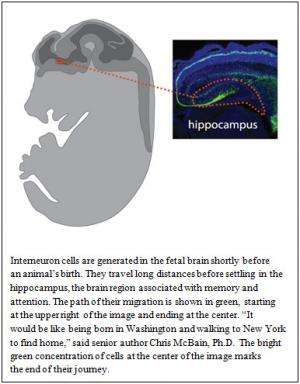
Key cells in the brain region known as the hippocampus are formed in the base of the brain late in fetal life and undertake a long journey before reaching their final destination in the center of the brain shortly after birth, according to researchers at the National Institutes of Health.
The hippocampus is involved with attention, navigation and converting short-term memories to long-term memories. Interneurons, the brain cell population the researchers studied, regulate communication between networks of brain cells. Previous research suggests that brain cell networks in the hippocampus may be disrupted in developmental disorders, including autism, as well as in epilepsy, Alzheimer’s disease and schizophrenia.
“The hippocampus seems to be at the crossroads of many disorders affecting the brain,” said Chris McBain, Ph.D., chief of the Laboratory of Cellular and Synaptic Neurophysiology at the NIH’s Eunice Kennedy Shriver National Institute of Child Health and Human Development (NICHD). “With these findings, we can begin to understand how proper communication is established in the brain and to investigate why sometimes it breaks down in this critical area.”
Their findings appear in The Journal of Neuroscience.
In their experiments, the researchers worked with two kinds of genetically altered mice. The researchers examined the brains of fetal mice and pups at various stages of development, from about two weeks after conception until the baby mice were 1 month old. Cells generated in the brains of the mice at each stage emitted light from a fluorescent protein they contained.
Comparing the number and location of fluorescent cells at the various stages, the researchers were able to create a composite image of the early development and migration of interneurons. The researchers also examined individual interneurons in the animals’ brains and catalogued nearly two dozen different types.
The researchers demonstrated that the brain produces large quantities of interneurons just before the mouse is born. Typically, brain cells reside in areas of the brain very close to where they are generated, Dr. McBain explained. But interneurons, the researchers discovered, take two to three days to travel across the brain to the hippocampus.
Along the route, most of the new brain cells die. Cell counts began to drop after the mice were born, the researchers found. When the mice were five days old, about half of the cells had disappeared. By the time the mice were one month old, the researchers estimated that 80 percent of the new interneurons had died.
The remaining cells were integrated into the hippocampus and demonstrated typical activity, the researchers showed. In the normally functioning brain, Dr. McBain explained, interneurons are incorporated in circuits connecting structures in different areas. In the circuit, they brake or sometimes stop signals at decisive moments. Working in concert with other cells in the circuit, they coordinate waves of activity across the brain.
“This work shows how the early development of the hippocampus is distinct from the rest of the brain,” said Dr. McBain. “It may also help us decide where to look when signals passing through somehow miss their mark.”


