Imaging the network traffic in our brains

MRI brain scans no longer just show the various regions of brain activity; nowadays the networks in the brain can now be imaged with ever greater precision. This will make functional MRI (fMRI) increasingly powerful in the coming years, leading to tools that can be used in cognitive neuroscience. This is the claim made by Prof. David Norris in his inaugural lecture as Professor of Neuroimaging at the University of Twente on 13 September.
During the twenty years since the invention of fMRI (functional Magnetic Resonance Imaging) developments have come thick and fast, from initially identifying active brain regions to more complex analysis of the connections and hubs in the brain. In his inaugural lecture Norris describes how this has been achieved thanks to not only a growing understanding of the underlying biophysics but also rapid technological developments: scanners with larger magnetic fields, better image-processing techniques and algorithms. His aim is to go beyond merely localizing which parts of the brain are active. The challenge is to answer two questions: How are the various regions interconnected, structurally and functionally? What do the networks in our brains look like?
Faster and more powerful
Back in the 19th century scientists observed increased blood flow in brain regions that are functionally active. fMRI enables the change in oxygen content to be seen. Haemoglobin, the substance that transports oxygen in the blood, can take the form of oxyhaemoglobin (when it is still combined with oxygen) and deoxyhaemoglobin (when the oxygen has been released), each of which has different magnetic properties. One of the complicating factors when interpreting the scans is that various physiological mechanisms are at work simultaneously, causing the deoxyhaemoglobin level to rise and fall. One of the remedies to increase accuracy, Norris explains, has been to increase the magnetic field strength: there are now MRI scanners operating at 7 Tesla. At the same time the speed at which laminae can be imaged has gone up by leaps and bounds: the entire brain can be scanned in three seconds with a precision of 1 millimetre.
Hubs
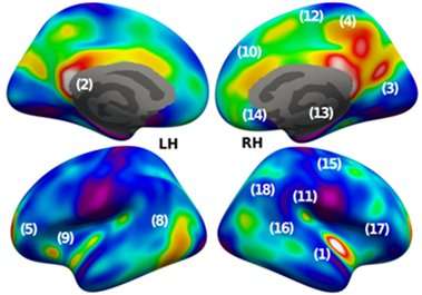
The functional connections between parts of the brain can be registered by means of blood flow, but MRI also enables the structural and anatomical connections to be seen. This involves measuring the movement of water molecules caused by the 'white matter' in nerve fibres. This technology is known as diffusion-weighted imaging (DWI). Combining these technologies provides a wealth of fresh information on the networks in the brain and the places where many connections come together, the 'hubs'. Not only have 'known networks' thus been proven, so have networks that neuroscience posits as plausible but that have never been measured.
More information: The new Centre for Medical Imaging that is to come to the University of Twente campus will soon provide extensive facilities for collaborating in the field of fMRI, says Norris, who is also on the staff of the Donders Institute in Nijmegen.


