Fetal heart ECG device breakthrough
Technology developed at The University of Nottingham has been used in a breakthrough study aimed at developing the first comprehensive model of a fully functioning fetal heart.
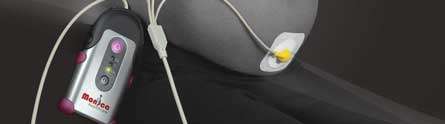
The abdominal fetal ECG device, designed originally by academics in the University's Department of Electrical and Electronic Engineering and on commercial sale throughout the world since 2008 through the University spin-out company Monica Healthcare Ltd, has been used to observe living fetal hearts of babies in their mothers' wombs.
The collaborative study led by experts at The University of Leeds has discovered that the walls of the human heart are a disorganised jumble of tissue until relatively late in pregnancy—with development much slower compared to other mammals.
Professor Barrie Hayes-Gill, Professor of Electronic Systems and Medical Devices at The University of Nottingham and joint founder and research director at Monica Healthcare said: "It's absolutely fantastic to see our device being used to detect fetal ECG morphology (i.e. ECG shape) in a non-invasive manner from the surface of the maternal abdomen. In this study the Monica device has been specifically deployed to observe the development of the fetal heart as it goes through gestation.
"It's an important development and we are delighted to see Nottingham technology playing such an integral part of the study. We expect that non-invasive morphological analysis during pregnancy and labour will become routine clinical practice in years to come as Monica continues to gain traction in the marketplace."
The fetal heart monitor is a portable, non-invasive device which attaches to the mother's abdomen and measures the electrical activity from the heart of the baby inside her womb. It is currently being used worldwide to monitor fetal heart rates during labour and delivery.
The device uses complex algorithms to correctly identify signals related to the fetal heart rate (FHR) using sensitive ECG-style electrodes. This method of using electrophysiological signals differs from current external monitoring devices that collect FHR and uterine activity data based on physical changes (e.g. change in reflected sound waves and changes on strain gauge) that may cause problems in data interpretation.
The monitor is simple to use, beltless, requires no wires to connect to the display or printer. There is also no need for the constant re-positioning of transducers, which is required with older technology and the mother is free to walk around if necessary.
As part of their study, which has been published in the Journal of the Royal Society Interface Focus, the team from the University of Leeds used the device to administer a weekly fetal ECG recording from 18 weeks until just before delivery.
The data from this, alongside two different MRI scans from the hearts of dead fetuses, was incorporated into a 3D computerised model built up using information about the structure, shape and size of the different components of the heart.
Early results suggest that the human heart may develop on a different timeline from other mammals. While the tissue in the walls of a pig heart develops a highly organised structure at a relatively early stage of a fetus' development, their work suggests there is little organisation in the human heart's cells until 20 weeks into pregnancy. Despite this, the human heart has a regular heartbeat from about 22 days.
Developing an accurate, computerised simulation of the fetal heart is critical to understanding normal heart developments in the womb and, eventually, to opening new ways of detecting and dealing with some functional abnormalities early in pregnancy.
Dr Eleftheria Pervolaraki, lead researcher on the project at the University of Leeds' School of Biomedical Sciences, said: "For a heart to be beating effectively, we thought you needed a smoothly changing orientation of the muscle cells through the walls of the heart chambers. Such an organisation is seen in the hearts of all healthy adult mammals.
"Fetal hearts in other mammals such as pigs, which we have been using as models, show such an organisation even early in gestation, with a smooth change in cell orientation going through the heart wall. But what we actually found is that such organisation was not detectable in the human fetus before 20 weeks," she said.
Professor Arun Holden, from The University of Leeds' School of Biomedical Sciences, said: "The development of the fetal human heart is on a totally different timeline, a slower timeline, from the model that was being used before. This upsets our assumptions and raises new questions. Since the wall of the heart is structurally disorganised, we might expect to find arrhythmias, which are a bad sign in an adult. It may well be that in the early stages of development of the heart arrhythmias are not necessarily pathological and that there is no need to panic if we find them. Alternatively, we could find that the disorganisation in the tissue does not actually lead to arrhythmia."
A detailed computer model of the activity and architecture of the developing heart will help make sense of the limited information doctors can obtain about the fetus using non-invasive monitoring of a pregnant woman.
Professor Holden said: "It is different from dealing with an adult, where you can look at the geometry of an individual's heart using MRI (Magnetic Resonance Imaging) or CT (Computerised Tomography) scans. You can't squirt x-rays at a fetus and we also currently tend to avoid MRI, so we need a model into which we can put the information we do have access to."
He added: "Effectively, at the moment, fetal ECGs are not really used. The textbooks descriptions of the development of the human heart are still founded on animal models and 19th century collections of abnormalities in museums. If you are trying to detect abnormal activity in fetal hearts, you are only talking about third trimester and postnatal care of premature babies. By looking at how the human heart actually develops in real life and creating a quantitative, descriptive model of its architecture and activity from the start of a pregnancy to birth, you are expanding electrocardiology into the fetus."


