Automated method could prevent blindness by detecting glaucoma in its early stages

A team of researchers led by Jun Cheng of the A*STAR Institute for Infocomm Research, Singapore, has developed a novel automated technology that screens for glaucoma more accurately and quickly than existing methods.
Glaucoma is a chronic, progressive eye disease that damages the optic nerve. It is the second leading cause of blindness worldwide, and will affect an estimated 80 million people by the year 2020. Progression of the disease can be slowed when treated early; however, glaucoma symptoms may go unnoticed until the advanced stages, by which time treatment is too late.
Currently, ophthalmologists use three methods to detect glaucoma. One is the assessment of increased pressure inside the eyeball. This method is not sensitive enough to detect glaucoma early and is not specific to the disease, which sometimes occurs without increased pressure. Another is the assessment of abnormal vision. This method requires specialized equipment, rendering it unsuitable for widespread screening.
The third method—assessment of the damage to the head of the optic nerve—is the most reliable but requires a trained professional and is time-consuming, expensive and highly subjective.
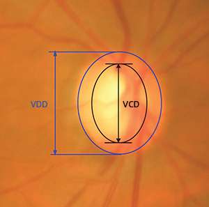
Glaucoma is characterized by a vertical elongation of the optic cup, a white area at the center of the optic nerve head, or optic disc. This elongation alters the cup-to-disc ratio (CDR) but does not normally affect vision. The computerized technique developed by Cheng and his colleagues measures the CDR from two-dimensional images of the back of the eye (see image).
The technique uses an algorithm that divides the images into hundreds of segments called superpixels and classifies each segment as part of either the optic cup or the optic disc. The cup and disc measurements can then be used to compute the CDR.
From 2326 test images, the researchers found that their automated technique is more accurate than the other glaucoma screening methods. Their technique takes around 10 seconds per image on a standard personal computer. This is comparable to other computerized methods, but automation makes it less laborious.
"The technique is ready to be used widely and can be used for screening so that glaucoma can be detected early," says Cheng. Early detection allows ophthalmologists to treat the patients, slowing disease progression.
Cheng also identifies potential improvements to the technique. "For example, we can include more data in the training to improve the accuracy." He says that the method can also be enhanced by integrating other factors, such as optic cup depth, into the analyses.
More information: Cheng, J., et al. Superpixel classification based optic disc and optic cup segmentation for glaucoma screening, IEEE Transactions on Medical Imaging 32, 1019–1032 (2013). ieeexplore.ieee.org/xpl/articl … jsp?arnumber=6464593


