September 25, 2013 report
Free-standing 3D skeletal muscle constructs created in the lab

(Medical Xpress)—Industrial robots can do incredible things, but their control systems are still incredibly complex. They rely largely on rotary electric power that is feedback-controlled, usually through precision optical encoders. Advances in artificial muscle technology, which could potentially simplify robot design, have been steady, but slow. With an eye towards building integrated bionic systems, researchers have been trying to construct devices based on real nerve and muscle. Most efforts so far have been limited to two-dimensional tissue culture systems which depend on adherence to a flat substrate for their integrity. Researchers at the University of Tokyo have recently fabricated free-standing, contractile muscle units from neural stem cells which they seeded on top of skeletal muscle. Their new paper, published in the journal Biomaterials, describes how these aligned fiber constructs can be stimulated to contract and generate significant force by chemical activation of the neurons—just like the real thing.
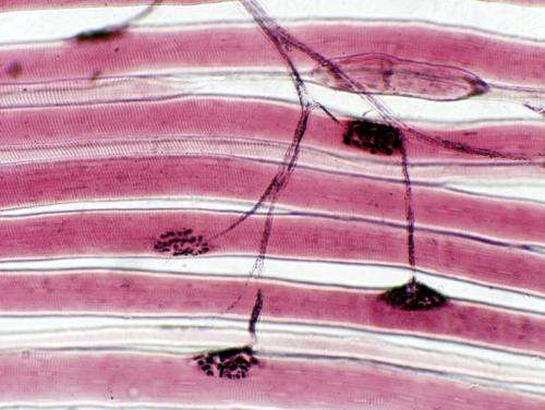
One essential step in growing realistic muscle is to create functional neuromuscular junctions (NMJs). In naturally grown muscle, each motorneuron generates around two thousand of these specialized synapses to control the contractive state of the fibers with its domain. The researchers found these NMJs in their constructs, and also the muscle-localized Acetylcholine receptors (ACh) that are needed to transduce control signals to the muscle. When they applied glutamate to activate the neurons, the muscles contracted in physiologic manner. In other words, the forces generated were uniformly aligned in a single direction across the tissue, and contracted in a synchronized fashion.
To fabricate aligned fiber bundles, the researchers used a polymer (PDMS) stamp to first create a striped pattern. The edges of the bundles were affixed to glass so that tension necessary for proper development could be maintained. The stem cell "neurospheres" were then added and differentiated into a mature form capable of extending axons into the muscle. During muscle development, or myogenesis, young muscle cells fuse into long multinucleated fibers known as myotubes. Not every cell survives this critical step and it is now known that certain cells most undergo apoptosis, or programmed cell death, for the tissue at large to pull this feat off.
The Tokyo researchers are not the only folks working in this field. A patent for a slightly different technique to build constructs with "functional interfaces" was granted just a couple years ago to researchers at the University of Michigan. This patent appears to include some provisioning for myotendinous attachments. As avascular tissues, tendons are a natural focus for replacement with engineered matrix materials. Integrating the natural nerve endings and sensors which transmit force and elongation will be critical to building actual functional devices.
As we recently reported, much remains unknown about exactly how NMJs are built and optimized by the organisms that use them. The manner in which the energy in the nerve pulse is partitioned into electrical and mechanical components as it propagates across the junction and into muscle is still much in debate. Nerve and muscle actuators, as ultimately envisioned here, may not be as robust as copper and steel—but for rebuilding or augmenting limbs, they are good materials to be familiar with.
More information: Three-dimensional neuron–muscle constructs with neuromuscular junctions, Biomaterials, Volume 34, Issue 37, December 2013, Pages 9413–9419. dx.doi.org/10.1016/j.biomaterials.2013.08.062
Abstract
This paper describes a fabrication method of muscle tissue constructs driven by neurotransmitters released from activated motor neurons. The constructs consist of three-dimensional (3D) free-standing skeletal muscle fibers co-cultured with motor neurons. We differentiated mouse neural stem cells (mNSCs) cultured on the skeletal muscle fibers into neurons that extend their processes into the muscle fibers. We found that acetylcholine receptors (AChRs) were formed at the connection between the muscle fibers and the neurons. The neuron–muscle constructs consist of highly aligned, long and matured muscle fibers that facilitate wide contractions of muscle fibers in a single direction. The contractions of the neuron–muscle construct were observed after glutamic acid activation of the neurons. The contraction was stopped by treatment with curare, an neuromuscular junction (NMJ) antagonist. These results indicate that our method succeeded in the formation of NMJs in the neuron–muscle constructs. The neuron–muscle construct system can potentially be used in pharmacokinetic assays related to NMJ disease therapies and in soft-robotic actuators.
© 2013 Medical Xpress


