New medical device performs surface imaging in surgery

Surgery is not for the faint of heart. Armed with a steady hand, extensive knowledge of human anatomy, and first-hand experience, a surgeon's task leaves very little room for error. Designed with a surgeon's mind and unique insight, a new breakthrough device developed by a team from Ryerson University gives surgeons a precise, 3D image of a surgical site's subsurface features, potentially increasing patient safety and saving time in the operating room, with direct impact on health care cost in hospitals.
Victor Yang, a Canada Research Chair in Bioengineering and Biophotonics at Ryerson University and a neurosurgeon at Sunnybrook Health Sciences Centre, remembers well the first few times he watched a master surgeon at work. Victor was amazed by the confidence displayed by the master surgeon on subsurface anatomy, almost as though he had X-ray vision. Judging from limited visible surface features and computing with "fuzzy logic", the master surgeon determined where and how to insert a screw into a spinal vertebral body, based on his knowledge of human anatomy and the patient's CT scans, yet without seeing what lay below the surface.
"There has to be a way to do that with a computer, with an imaging system," Yang recalls thinking. "We should be able to measure that, precisely, to sub millimeter level and as fast, if not faster, than the subconscious fuzzy logic previously used by surgeons."
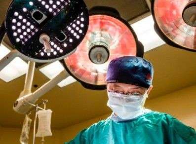
The 7D Surgical Navigation device was borne out of the research that followed at the Biophotonics and Bioengineering Lab (BBL). The device, which looks like an ordinary operating room light filled with LED (light emitting diodes), shines a sequence of light onto the surgically exposed surface at very high speed. The computer then captures the light that reflects off of the surgical site's surface and calculates its three-dimensional shape, called topographical imaging. The computer matches the surface image information with what is captured by the patient's MRI/CT scan. The result is the ability to visualize the entire surgical site in real-time, including subsurface information, the likes of which has never been available to surgeons.
Currently being tested at Sunnybrook Health Sciences Centre, the time saved with the new device is impressive. The 7D Surgical Navigation system performs at least 10 times faster than existing systems; the device takes 1-2 minutes to capture and compute the information needed to begin surgery, while similar products take 20 minutes or more. In the operating room, each minute counts, anywhere from $100-200. In addition to cost savings, shorter times on the operating table are also better for patients.
7D Surgical is now a spin-off company comprised of Yang's research team members at Ryerson University, in partnership with Sunnybrook Research Institute. As the co-founder of 7D Surgical and chief scientific officer, Yang started the conception designs and led the team through bench testing, pre-clinical, and finally clinical demonstration. The BBL team co-founders include Beau Standish, chief executive officer of 7D Surgical, business development; Adrian Mariampillai, chief technology officer, optics and algorithm design; Michael Leung, lead programmer, software development; David Cadotte, clinical research scientist, surgical technique development; and Peter Siegler, senior research scientist, hardware and surgical tools.
The project, which began in 2010, has been funded by a number of sources including MaRS Innovation, Natural Sciences and Engineering Research Council of Canada, Canadian Institutes of Health Research, Canada Research Chairs program, Canada Foundation for Innovation, Ontario Brain Institute, and the Networks of Centres of Excellence.
Clinical testing on the device began this spring and so far, says Yang, feedback from surgeons at Sunnybrook has been extremely positive, or, more pointedly: "When do we get to start using this?"
According to Yang, also a senior scientist at Sunnybrook Research Institute, "Preliminary results show that the technology can perform surface imaging in a real surgical environment with all the interferences of a normal operating room and we are able to lock on to target and show subsurface anatomy to the attending surgeons."
The full trial will include 60 patients and will be tested in both spinal and brain surgeries. Yang and his team expect that the full trial will be completed within the year.
While there is currently only one unit in use at Sunnybrook, a new partnership with Celestica, a manufacturing company that specializes in accelerated manufacturing, is changing that. Within the year, Yang and the 7D Surgical team predict that five additional units will be made and placed at additional clinical sites. The goal is to have the device in operating rooms around the world.


