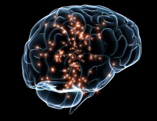
Lasers, magnetism allow glimpses of the human brain at work

To the untrained eye, it looked like a seismograph recording of a violent earthquake or the gyrations of a very volatile day on Wall Street—jagged peaks and valleys in red, blue and green, displayed on a wall. But the story it told was not about geology or economics.
It was a glimpse into the brains of Shaul Yahil and Shaw Bronner, two researchers at a Yale lab, as they had a little chat.
"This is a fork," Yahil observed, describing the image on his computer. "A fork is something you use to stab food while you're eating it. Common piece of cutlery in the West."
"It doesn't look like a real fancy sterling silver fork, but very useful," Bronner responded. And then she described her own screen: "This looks like a baby chimpanzee ..."
The jagged, multicolored images depicted what was going on in the two researchers' heads—two brains in conversation, carrying out an intricate dance of internal activity. This is no parlor trick. The brain-tracking technology at work is just a small part of the quest to answer abiding questions about the workings of a three-pound chunk of fatty tissue with the consistency of cold porridge.
How does this collection of nearly 100 billion densely packed nerve cells, acting through circuits with maybe 100 trillion connections, let us think, feel, act and perceive our world? How does this complex machine go wrong and make people depressed, or delusional, or demented? What can be done about that?
These are the kinds of questions that spurred President Barack Obama to launch the BRAIN initiative in 2013. Its aim: to spur development of new tools to investigate the brain. Europe and Japan are also pursuing major efforts in brain research.
The mysteries of this organ, which sucks up about 20 percent of the body's energy, are many and profound. But with a collection of sophisticated devices, scientists are peering inside the working brains of people for clues to what makes us tick.
After all, while a lot can be learned from dissecting brains after autopsy or studying animal brains, there's nothing quite like watching a human one work. A brain is like a car motor, says researcher Joy Hirsch. You can study it at rest, but "until you start that car up and run it, you don't really see how all the working parts work together and what the dynamics are."
The Yale Brain Function Lab, which she directs, is investigating how our brains let us engage with other people. That's one of the most basic questions in neuroscience, as well as an ability impaired in autism and schizophrenia, she said: "It's probably one of the most fundamental functions of the human species, and yet we know very little about it."
Hirsch turned to a decades-old brain mapping technique that only recently has developed far enough for her task.
As Yahil and Bronner chatted to demonstrate the technology, each wore a black-and-white skullcap from which 64 slender black cables trailed away like dreadlocks. At the tip of half of those fiber optic cables, weak laser beams slipped through their skulls and penetrated about an inch into their brains. There, the beams bounced off blood and reflected back to be picked up by the other half of the cables.
Those reflections revealed how much oxygen that blood was carrying. And since brain circuits use more oxygen when they're busier, the measurements provided an indirect index to patterns of brain activity as Bronner listened to Yahil and replied, and vice versa.
The technique monitors only areas close to the brain's surface, and so misses signals from its emotional centers, for example. But it does show, for example, that watching a conversational partner's face makes a difference in what brain circuits turn on during a chat.
Using a similar setup, researchers in China recently created groups of three college students who were given a topic and told to discuss it. Results showed that students who emerged as leaders showed greater synchronization of brain patterns with their followers than pairs of followers did. The better the leader's communication skills, the more profound this link was.
Such studies are unusual in that they monitor multiple brains simultaneously. Usually, experiments examine just one at a time.
Researchers have caught glimpses of human brain activity since the 1920s, when scalp electrodes first eavesdropped on the electrical chatter of brain cells to produce the zig-zag lines of EEG. Early images appeared in the late 1970s and 1980s through other techniques, like PET scans.
But the coming-out party for the brain-mapping technique most widely used today came at a 1991 meeting in San Francisco. Scientists watched what one observer called a "jaw dropping" movie that showed activation of a part of the brain that handles visual information. That movie was created through functional magnetic resonance imaging, or fMRI.
Basically, fMRI does what Hirsch's laser system does: It uses oxygen levels in blood as tracers of brain-cell activity. But it penetrates much deeper into the brain, using powerful magnetic fields. That lets it seek subtle magnetic signals to track blood oxygen levels on a tiny scale; a bump in oxygen levels indicates active brain cells nearby.
The result is visually striking: detailed brain images with bright blotches of color that indicate where the action is. That's not a direct picture, like an X-ray showing a broken bone, but rather a reconstruction that involves some key assumptions and a lot of sophisticated number-crunching.
How sophisticated? Consider the researchers who put a four-pound dead Atlantic salmon in an MRI scanner, and asked it to determine what emotions were being experienced by people in some photos. It sounds absurd, but some raw results indicated brain activity in response. The lesson, the researchers said, is to be careful about statistical analysis of the complicated data that goes into producing brain images.
Even when that's done right, fMRI is not perfect. It usually samples the brain less than once per second, which is glacial compared to the rapid fire of activity. And while its resolution is high, with data coming from cube-shaped volumes a few hundredths of an inch on a side, each cube can still contain hundreds of thousands of brain cells. If a cube shows up as active, you can't tell which of its cells are working.
And if fMRI shows two brain regions to be active at the same time, it can't reveal which one is turning on the other.
Still, fMRI can detect changes in brain activity that are associated with particular tasks, and they are vanishingly tiny. You may think you are busting your brain to solve a difficult crossword puzzle or recall somebody's name. But such activities make only a tiny difference in the brain's overall energy consumption, says Dr. Marcus Raichle of the Washington University School of Medicine.
That's because "most of what your brain is doing, it's doing it all the time," Raichle said.
So what are our brain circuits doing when we aren't focused on anything?
That is "probably one of the most researched topics right now" in brain mapping, says John Darrell Van Horn of the University of Southern California.
Brain scans show that when people are at rest, without a particular mental task to perform, their brains continue to hum along as particular regions communicate with each other. Research suggests that more often than not, people are thinking about humdrum matters like taking the car for an oil change or a bothersome conversation, Van Horn said.
Scientists are studying what these patterns of brain activity at rest can reveal about the brain and its illnesses. Altered patterns have shown up in such conditions as panic disorder and depression. Older adults show less activity than younger people in one particular pattern, and the decline seems more pronounced in people with Alzheimer's disease, said Mara Mather, professor of gerontology and psychology at USC.
People with autism also show less activity in that network, she said.
And people with schizophrenia also show different patterns in the resting state than healthy people do, says Peter Bandettini of the NIH. That lets researchers cast a wide net in looking for specific differences in brain function, without having to search for specific mental tasks to assign people to do in order to make the differences apparent, he said.
While the brain at rest is not completely understood, "everyone is jumping into this now," Bandettini said. It holds promise for mapping out which parts of the brain work with which others to perform key tasks, he said.
The emphasis in brain mapping these days is not so much about finding particular places that do particular tasks, but rather delineating the circuitry that lets the brain operate.
"No region works in isolation. It's all being communicated across networks," Mather said.
Communication flows along an estimated 150,000 miles of nerve fibers in the average brain. Individual fibers are too fine to see in brain-scanning machines, but they form bundles that can be detected as they cross the deep central portion of the brain.
Those bundles are one focus of researchers who are mapping out the brain's "connectome," the complex web of these connections between areas of gray matter, where thinking takes place.
"Any one patch of gray matter in the brain is literally communicating with hundreds of other distant locations, in ways that actually are very different from how modern computer circuits are wired," says David Van Essen of Washington University.
While computers use their lightning speed to crunch numbers, the brain's "squishy hardware," works with far slower internal communication, he said.
"We're able to do analyses and make inferences in ways that still way outperform computers, because the wiring is different and in some ways just incredibly clever. But we don't understand the rules, the strategies in sufficient detail."
The connectome effort is still in its early phases, he said. But it has already gained one unusual distinction: A colorful scientific depiction of brain connections made the cover of an album by the band Muse.
Beyond that, one aspect is mapping out what parcels of tissue do what jobs in the brain's outer layer, the cerebral cortex. The maps that show up in textbooks are about as sophisticated as 16th-century maps of the Earth, Van Essen said.
"What we know now but isn't yet in the textbooks is more like a 17th century map ... reasonably accurate in some places and humorously off track in the so-called unexplored continents," he said.
The Human Connectome project should bring the maps up to the equivalent of 18th- or 19th-century Earth maps, he said.
That's a long way from Google-like precision.
"Navigating your brain or my brain down the level of this neuron or that neuron as we are thinking and talking, some of those things are way out of scope for what we can do or even predict," Van Essen said. "Some of us (think) that may be beyond what the human brain can ever achieve."
Some brain-scanning research rises from the informative to the truly startling, like decoding—looking at brain activity patterns to figure out what somebody is seeing, or even thinking about.
In 2011, for example, researchers reported that they could reconstruct very rough visual replicas of movie clips that people were watching while their brains were scanned. And two years later, Japanese scientists reported evidence that they could get some idea of what people were dreaming about—at least, better than chance under highly controlled conditions.
Such findings are valuable for learning how the brain is organized. And in the near term, decoding technology might help people whose medical condition prevents normal conversation, said Jack Gallant of the University of California, Berkeley.
As brain-scanning machines and know-how progress, scientists expect more discoveries. Already, a technology that lets MRI machines capture brain images several times a second rather than less than once per second is becoming more widely used, Van Horn said. It's already clear that the faster sampling provides new information, and scientists are just beginning to understand how to interpret it, he said.
And while fMRI can't reveal now whether a particular activated area is stimulating another one, that may change in the future, Bandettini said. The key advances would be if scientists can better measure tiny lags in activity between different places in the brain, or get higher resolution images that distinguish between brain cells that are sending signals and those that are receiving them.
Hirsch, meanwhile, is testing whether the monitoring technology she uses to track two brains in conversation can be made portable. If so, she could move her experiments out of the lab and into more natural settings.
"It would be easier to image people when they're walking and talking, or exercising or walking down an aisle in a grocery store, or making decisions on the fly—driving a car, for example," she says.
Making portable devices to replace huge MRI machines could open new possibilities for brain decoding. And not just for scientists.
Gallant foresees a future in which composers write music just by imagining it. Or "you can just think about the picture you want to paint" and let a computer do the rest.
Writing a letter, he says "would be like dictation, except you would just be talking to yourself."
And in the future, why be confined to your own language?
"I can think in English and my little brain hat would read my thoughts, send it to Google and it would come back in Japanese," he says. "You'd talk out of a little speaker in your hat."
"There's nothing theoretically challenging about that. It's just a matter of getting good enough data."
More information: BRAIN initiative: www.whitehouse.gov/brain
European brain project: www.humanbrainproject.eu
Japanese brain project: brainminds.jp/en
© 2015 The Associated Press. All rights reserved.


