Detecting melanoma early, without a biopsy

Melanoma is a form of skin cancer that becomes dangerous when it spreads, but is treatable in its early stages. Doctors diagnose melanoma by cutting away a piece of a suspicious skin lesion—a procedure known as a biopsy—and testing it for malignant cells.
It's an imperfect, invasive method that Colorado State University researcher Jesse Wilson wants to improve. His goal is to make early detection of melanoma faster, cheaper and less invasive than a biopsy.
An assistant professor in the Department of Electrical and Computer Engineering and in the School of Biomedical Engineering, Wilson's expertise is in pushing the boundaries of biomedical optics. He has received a one-year, $30,000 grant from the Colorado Clinical and Translational Sciences Institute to develop a new microscope that can distinguish between benign and malignant pigmented skin lesions, without the need for biopsy.
If his idea works, it could lead to low-cost, in-vivo imaging of melanin, the skin pigment that's made by cells called melanocytes, which can become cancerous and lead to melanoma.
The grant is through the CCTSI's "CO-Pilot" program, which provides start-up funds for early-career researchers embarking on multidisciplinary new ideas. Wilson, who did his undergraduate, master's and Ph.D. work at CSU, has returned to campus as a faculty member to continue applying cutting-edge biomedical optics to early cancer detection.
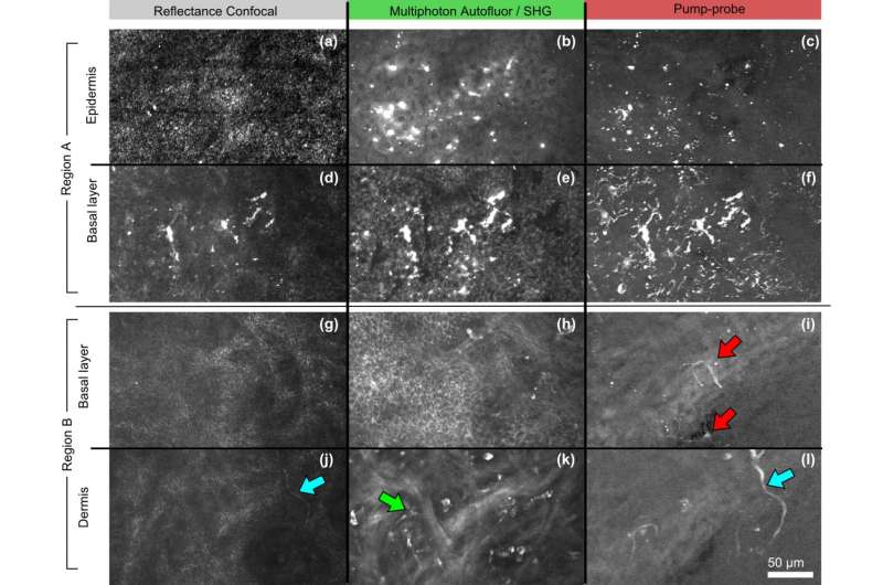
The CO-Pilot is funding Wilson's development of an experimental microscope that uses a technology called pump-probe, which can provide contrast between normal tissue and melanoma without stains or dyes.
Pump-probe is a type of multiphoton microscopy, an established technique used for deep imaging of living tissue. In standard multiphoton microscopy, an extremely short laser pulse is used to excite fluorescent molecules in the sample, which can then be detected when they light up.
In pump-probe, excited molecules are detected not through fluorescence, but through their interactions with a second laser pulse. This allows pump-probe to distinguish molecules based on their absorption properties, which provide strong contrast between different types of melanin.
Pump-probe microscopes that can detect melanoma were developed in the lab of Warren Warren at Duke University, where Wilson completed his postdoctoral training. However, these microscopes require a short-pulse laser source that costs upwards of $300,000 - a major barrier to commercializing the technology for widespread use.
That's the problem Wilson will tackle with the CO-Pilot grant. He is designing a pump-probe microscope that can distinguish between benign and malignant melanoma in vivo, but built around a simpler laser source that's already widely used in telecommunications applications to encrypt voice communications. That laser only costs about $5,000, and would make the pump-probe technology more realistic for melanoma applications.
Wilson will work with Ali Pezeshki, associate professor in ECE and mathematics, on the signal processing that will be central to constructing images with the new microscope. He also plans to collaborate with Dan Gustafson, director of research at the Flint Animal Cancer Center.
Wilson's lab is also pursuing other pump-probe applications, including imaging cytochromes, molecules involved in metabolic activities that can't be imaged with conventional techniques.


