'Fixing' blood vessel cells to diagnose blood clotting disorders

When in dysfunction, the vascular endothelium—the tissue that lines the blood vessels throughout our body's entire circulatory system—plays a big role in the development of many human diseases, including diabetes, stroke, heart disease, viral infections and cancer. This is because endothelial cells are sensitive to blood flow and also interact with blood cells through molecules on their surface, so that blood coagulation and platelet function are modulated. In normal 'hemostasis', the endothelium prevents deadly blood loss and clot formation. However, dysfunction or inflammation of the endothelium may result in aberrant blood coagulation inside the vessels, leading to life-threatening blockages or hemorrhage.
"Abnormal blood coagulation and platelet activation are major medical problems and the ways we study them now are overly simplified," said Wyss Institute Founding Director Donald Ingber, M.D., Ph.D., who is also the Judah Folkman Professor of Vascular Biology at Harvard Medical School and the Vascular Biology Program at Boston Children's Hospital, and Professor of Bioengineering at the Harvard John A. Paulson School of Engineering and Applied Sciences. "Clinicians currently do not have tools to monitor hemostasis that take into account physiologically-important interactions between endothelial cells and flowing blood."
Until now, the crucial interface between endothelial cells and circulating blood has not been accurately replicated in a practical diagnostic device, due to the challenge of incorporating living endothelial cells into a robust testing tool.
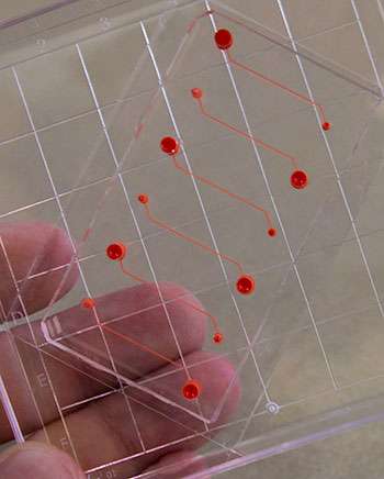
Now, a team led by Ingber at the Wyss Institute for Biologically Inspired Engineering at Harvard University has discovered that endothelial cells need not be 'living' in order to confer their effects on blood coagulation. A new device developed by the team, published in August in the journal Biomedical Microdevices, could monitor blood clot formation and diagnose effectiveness of anti-platelet therapy, by microengineering tiny hollow channels lined by chemically 'fixed' human endothelial cells that more closely mimic cellular and vascular flow conditions inside a patient's body than a bare surface.

"It's a bioinspired device that contains the endothelial function of a diseased patient without having actual living cells, and this greatly increases the robustness of the device," said the study's first author Abhishek Jain, Ph.D., a former Wyss Institute Postdoctoral Fellow who has recently been appointed Assistant Professor of Biomedical Engineering at Texas A&M University.
This blood coagulation diagnostic can even be used to study the effects of endothelial inflammation on the formation of blood clots, which is highly relevant in patients suffering from atherosclerosis, a chronic disease that builds up plaque leading to hardening and narrowing of blood vessels.
"This is one of the first examples of how a microfluidic cell culture system could have added value in clinical diagnostics," said study co-author Andries van der Meer, Ph.D., a former Wyss Institute Postdoctoral Fellow who is now Assistant Professor in Applied Stem Cell Technologies at University of Twente, The Netherlands. "Using chemically fixed tissue that is no longer alive offers a clear, low-risk path toward further testing and product development."
A previous study by Ingber and his team showed that recreating the physicality and blood flow of vasculature within microfluidic channels allowed them to predict precise times that blood might clot, with potential applications in real-time monitoring of patients receiving intravenous anticoagulants in order to prevent complications such as stroke and vascular occlusion. Their latest device adds another layer of complexity by embedding the functionality of the vascular endothelium within a diagnostic tool that might be manufactured, stored and shipped for clinical use, which was never before considered possible.
"Our efforts to mimic the vascular system in a meaningful way within a microfluidic device has led to two avenues of technology development, which could potentially be combined in the future to develop portable tools suited for diagnosing and even discovering what disease states lead to blood clotting," said Ingber. "Together they represent a new suite of physiologically-relevant microdevices that incorporate critical mechanical cues, and which could have near-term impact on our understanding and prevention of dysfunctional hemostasis."
More information: Abhishek Jain et al. Assessment of whole blood thrombosis in a microfluidic device lined by fixed human endothelium, Biomedical Microdevices (2016). DOI: 10.1007/s10544-016-0095-6


