Oligodendrocytes selectively myelinate a particular set of axons in the white matter

There are three kinds of glial cells in the brain: oligodendrocytes, astrocytes and microglia. Oligodendrocytes myelinate neuronal axons to increase conduction velocity of neuronal impulses. A Japanese research team at the National Institute for Physiological Sciences (NIPS, Okazaki, Aichi, Japan) has found a characteristic feature of oligodendrocytes that selectively myelinate a particular set of neuronal axons.
It is known that maturation of oligodendrocytes is necessary for motor skill learning. The structure of the white matter changes after motor skill training (e.g., juggling or playing piano). These reports suggest that a single oligodendrocyte selectively myelinates a particular set of axons. In addition, oligodendrocyte dysfunction causes severe neurological disorders, such as multiple sclerosis. So better understanding of the interactions between oligodendrocytes and neuronal axons has important medical applications. However, the difficulty of identifying the interaction between oligodendrocytes and neuronal axons in the brain was due to the high density of oligodendrocytes in white matter, preventing researchers from detecting the precise morphology of each oligodendrocyte.
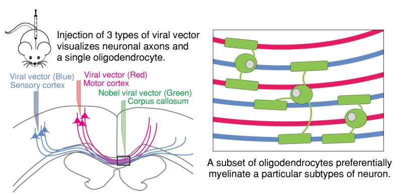
The Japanese research group at NIPS used a viral vector to label single oligodendrocytes in the white matter. With multiple viral vector injections, neuronal axons derived from distinct brain region (motor cortex or sensory cortex) and oligodendrocytes in the white matter were simultaneously labeled. Surprisingly, the research group found that oligodendrocytes did not just ensheath axons randomly—some oligodendrocytes selectively myelinated axons from a particular brain region.
This method developed by the research group is available for demyelination in an animal model to assess demyelinating diseases. "Now, we plan to analyze oligodendrocyte morphology and myelination in demyelinating mouse models," says corresponding author Dr. Shimizu. "Furthermore, axon selective myelination for a specific neuronal subtype found in this study encourages us to investigate physiological relevance of multiple myelination to higher brain function."
More information: Yasuyuki Osanai et al, Rabies virus-mediated oligodendrocyte labeling reveals a single oligodendrocyte myelinates axons from distinct brain regions, Glia (2016). DOI: 10.1002/glia.23076


