January 9, 2017 report
Partially developed brain found in young girl's ovary

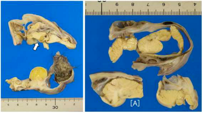
(MedicalXpress)—A team of surgeons performing a routine appendectomy on a young woman also found and removed a tumor they noticed growing on their patient's ovary. Subsequent analysis of the tumor by researchers with the Shiga Medical Centre for Adults in Japan revealed that the tumor contained a teratoma with a brain-like structure along with a partially developed skull bone and hair fragments. They have published their findings in the journal Neuropathology.
Teratomas, also known as dermoid cysts, are not all that uncommon, the researchers report, but ones that have brain-like structures are extremely rare. In this case, the teratoma, which is Greek for the word monster, held a brain-like structure that was so advanced it had partially developed into a cerebellum with a brain stem and was able to transmit electrical pulses delivered by the research team.
The tumor was approximately 10 centimeters wide and held what the researchers describe as a mat of greasy hair and a brain-like structure that was covered by skull-like bone material. Teratomas are actually a type of tumor that develop most often in organs such as the thyroid, liver, lung, brain and ovaries—their defining characteristic is the development of bodily material that is out of character for the location in which it is found. Why they occur is not known, but prior research has suggested that, like cancerous tumors, they result from a malfunction during cell division, often in ways that resemble abnormal stem-cell growth. The researchers also note that approximately 20 percent of ovarian tumors have teratomas, which often have hair, teeth, muscle, fat or cartilage and of course sometimes brain cells. Very seldom are such cells organized into anything resembling brain parts.
In some cases, such tumors can cause symptoms due to the immune system being activated, but the patient in this case, a 16-year-old girl, had no symptoms before her surgery and recovered quickly after removal of both appendix and tumor, the researchers note. They also observe that despite the uneasiness some may feel regarding teratomas, they offer researchers insights into human development that can be found no other way.
More information: Masayuki Shintaku et al. Well-formed cerebellum and brainstem-like structures in a mature ovarian teratoma: Neuropathological observations, Neuropathology (2017). DOI: 10.1111/neup.12360
Abstract
In the surgical case of a mature cystic teratoma of the ovary that arose in a 16-year-old girl, a large amount of well-differentiated and highly organized cerebellar tissue was found. Three layers of the cerebellar cortex were well formed, and synaptophysin-positive "glomeruli" were found in the granule cell layer. Some Purkinje cells exhibited focal expansion and a dysmorphic appearance of the dendrites. Adjacent to the cerebellar tissue, a large space lined by the ependymal layer and a club-shaped CNS tissue mass resembling the brainstem were found, and structures reminiscent of the midbrain tectum and pontine nuclei were distinguished within this mass. The CNS tissue was surrounded by the leptomeninges and a skull-like, bony shell. Structures consistent with the supra-tentorial CNS tissue were not found. This case represents an example of infra-tentorial CNS tissue that was well-differentiated and organized to an exceptionally high degree in an ovarian mature teratoma. Various degenerative changes have been documented in CNS tissue in ovarian teratomas, but the dendritic abnormalities of Purkinje cells seen in the present case are novel findings.
© 2017 MedicalXpress


