October 12, 2017 report
A portable tiny brain scanner for studying brain disorders in infants
")
(Medical Xpress)—A team of researchers affiliated with several institutions in France has developed a new type of brain scanner that is small enough for use on infants. In their paper published in the journal Science Translational Medicine, the group describes the new device, how it was tested and its effectiveness in initial trials.
When studying brain disorders that cause seizures, such as epilepsy, researchers have typically used EEG and fMRI to study the electrical signals that are generated by the brain and the regions of the brain that are impacted. But to learn more, researchers would like to be able to trace the progression of such disorders from an earlier age—ideally, they would like to be able to start right after a baby is born. But EEG requires gluing probes to the skull and fMRI machines are big, loud and scary, so infants have gone without such testing. But now, that appears likely to change, as the researchers with this new effort have built a machine that is small enough to carry (it weighs just 40 grams), is quiet, and can be used even on sleeping infants.
The new machine, called fUSI (functional ultrasound imaging) is small enough to place in a crib—it connects to a cap that looks much like the knitted caps often used in hospitals to keep infants warm. The machine combines video-electroencephalography and ultrasound to map out blood flow related to changes in electrical signals in the brain and actually offers higher resolution than conventional machines.
")
The team reports that the machine has been tested on healthy infants (with no brain disorder) and on two infants with drug-resistant seizure disorders. They found that the machine was able to detect neurovascular changes during seizures in the cortex and then to trace them back to their source. The machine captures brain imagery continuously, the team notes, making it ideal for situations where abnormal brain activity occurs at unpredictable moments. The group believes their new machine will very soon become standard equipment for bedside monitoring of infants with brain disorders and will provide a new tool for researchers looking to understand the root causes of brain disorders and to ultimately prevent them from developing.
-
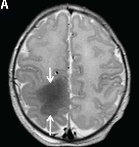
MRI brain scan of a patient with cortical dysplasia. Credit: C. Demene et al., Science Translational Medicine (2017) -
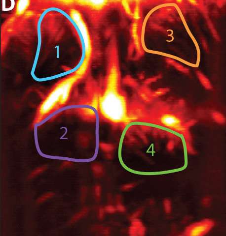
Representative brain image from an infant. Credit: C. Demene et al., Science Translational Medicine (2017) -
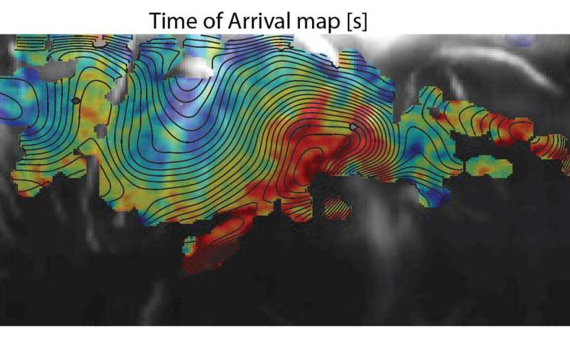
Data from a new noninvasive imaging method for monitoring brain activity in infants. Credit: C. Demene et al., Science Translational Medicine (2017)
More information: Charlie Demene et al. Functional ultrasound imaging of brain activity in human newborns, Science Translational Medicine (2017). DOI: 10.1126/scitranslmed.aah6756
Abstract
Functional neuroimaging modalities are crucial for understanding brain function, but their clinical use is challenging. Recently, the use of ultrasonic plane waves transmitted at ultrafast frame rates was shown to allow for the spatiotemporal identification of brain activation through neurovascular coupling in rodents. Using a customized flexible and noninvasive headmount, we demonstrate in human neonates that real-time functional ultrasound imaging (fUSI) is feasible by combining simultaneous continuous video–electroencephalography (EEG) recording and ultrafast Doppler (UfD) imaging of the brain microvasculature. fUSI detected very small cerebral blood volume variations in the brains of neonates that closely correlated with two different sleep states defined by EEG recordings. fUSI was also used to assess brain activity in two neonates with congenital abnormal cortical development enabling elucidation of the dynamics of neonatal seizures with high spatiotemporal resolution (200 μm for UfD and 1 ms for EEG). fUSI was then applied to track how waves of vascular changes were propagated during interictal periods and to determine the ictal foci of the seizures. Imaging the human brain with fUSI enables high-resolution identification of brain activation through neurovascular coupling and may provide new insights into seizure analysis and the monitoring of brain function.
© 2017 Medical Xpress


