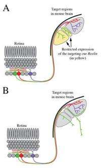
A. Schematic representation of axonal connections between different classes of retinal neurons (colored in green, red or purple) and target regions within the brain. Note that the axons from these three neurons branch into different brain regions. Two target regions depicted here contain the targeting cue Reelin, color coded in yellow. B. In mice lacking reelin protein, the axonal projections of one of these classes of neurons (colored in green) are mistargeted into inappropriate brain regions. Note that axons from the other classes of retinal neurons are unaffected by the absence of reelin. Image courtesy of Michael A. Fox, Ph.D./VCU.
(PhysOrg.com) -- Virginia Commonwealth University School of Medicine researchers have identified a mechanism that regulates the formation of a specific neural circuit, taking researchers a step closer to understanding how the human brain works.
By studying the visual system, Michael A. Fox, Ph.D., assistant professor in the Department of Anatomy and Neurobiology, and his research team have been working to create a road map for classes of neurons in the retina – a challenging task despite the breadth of research focused on circuit formation in the visual system.
The human brain has billions of neurons that connect with each other in very precise, stereotyped patterns. The network of connections between specific groups of neurons, called neural circuits, are responsible for very specific behaviors, such as our ability to sense the environment, to move or even to assemble memories. For this reason, it is critical that these neural circuits be correctly generated during brain development.
According to Fox, with billions of neurons, and each capable of generating thousands of connections, it is often extremely difficult to identify the cues that allow the correct partner neurons to find each other and form lasting connections, called synapses.
Now, in a new study published online in the Jan. 12 issue of The Journal of Neuroscience, Fox and his team report a mechanism by which a set of axons can target a correct region of the brain, and therefore form a precise, specific neural circuit. Specifically, the team identified a cue called reelin that was necessary for axons from one class of retinal neurons to target the correct region of the brain.
“To date, this is the first cue to be involved in what we call class-specific targeting of axons in the visual system,” said Fox.
“These findings may also provide novel clues into how parts of the brain function - to understand brain function we have to know which neurons are connected into circuits. Equally important, our findings may also lay the foundation for regenerative studies,” he said.
Using a mouse model, the team compared molecules in regions of the brain that were conducive to the growth of axons from some classes of retinal neurons against adjacent regions that inhibited axonal growth.
According to Fox, there are a number of diseases, such as glaucoma, that affect neurons in the retina and lead to visual impairment or even blindness. In glaucoma, pressure buildup in the eye damages and kills the retinal neurons that connect with targets in the brain.
Researchers are investigating ways to potentially cure glaucoma and related diseases by replacing dead or diseased cells with stem cells.
“While much progress has been made in the efforts to harness the pluripotent capacity of stem cells, to incorporate stem cells into diseased tissue, and to induce them to differentiate into the correct types of neurons, we still need to know how to get the new retinal neurons to incorporate into correct neural circuits,” said Fox.
“This means we will need to know exactly how each neuron finds its appropriate target neuron. We are a long way from knowing just how to do this, but our study moves us one step closer,” he said.
Next for Fox and his team will be to use some of the data collected to further explore the role of other cues in the targeting of other classes of these axons. Eventually, they hope to learn exactly how each axon finds the right target region or neuron in the brain.
Provided by Virginia Commonwealth University


