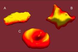
Deoxygenated red blood cells (A, B) from patients with sickle cell disease have a highly irregular morphology, compared to the characteristic biconcave shape of normal erythrocytes (C). These three-dimensional topographical images were created via an atomic force microscope by graduate student Jamie Maciaszek.
Until recently, the pairing of molecular biology and mechanical engineering would have been viewed as highly unusual. But thanks to an explosion of imaging and simulation techniques over the past few decades, the opportunity now exists to view biology at the molecular and even nano-levels through the eyes of an engineer.
George Lykotrafitis, an assistant professor in the Department of Mechanical Engineering, is making full use of this opportunity. Using state-of-the-art instruments, Lykotrafitis is researching the intricacies of sickle cell anemia, a painful, debilitating disease affecting one in every 5,000 Americans, that unleashes numerous health complications and reduces by half the life expectancy of those who are afflicted. The rate is even higher in developing regions of the world.
Sickle cell anemia, or sickle cell disease, involves a genetic mutation that alters the genetic code of hemoglobin, a protein on red blood cells tasked with carrying oxygen throughout the body. Normally, red blood cells are highly elastic and able to alter their shape to pass easily through capillaries, but mutated hemoglobin proteins link together to form a network of stiff polymer fibers. These long fibers distort the shape, strength, and elasticity of the cell, and along with increased red blood cell adherence to the blood vessels make it difficult for red blood cells to squeeze through tiny capillaries, often producing vessel blockages or tears. This can reduce oxygen flow, which can severely damage organs and cause intense pain if left unchecked.
In addition, the defining sickle shape of the red blood cells decreases the surface area to which oxygen can bind, increasing oxygen deprivation. And the lifespan of sickle cells is much less than that of normal red blood cells, resulting in anemia and further reduction in oxygen flow throughout the body.
In studying sickle cell anemia, Lykotrafitis aims to quantify the different characteristics of the membranes of erythrocytes with sickle cell anemia. He is particularly interested in studying the elasticity of the cell membrane and the presence of certain proteins on the cell membrane that encourage adhesion to proteins on the inner lining of blood vessels. Using various tests to study these characteristics, he is able to advance understanding of the disease.
“What we do here, basically, is quantify,” he says. “Then with that baseline, we can see how drugs affect specific cells.”
A critical tool Lykotrafitis employs in examining cell membranes is atomic force microscopy (AFM). In contrast with a standard optical microscope, which uses light to illuminate a surface, an atomic force microscope “feels” the surface and enables researchers to measure the texture and elasticity of the membrane.
In AFM, an extremely small needle moves systematically up and down along the entire surface it is scanning, recording the texture of the surface by assigning the specific position on the sample to a corresponding “altitude” and creating a kind of topographic map of the surface that shows the shape and texture of the membrane. This provides a detailed, three-dimensional image that cannot be produced with an optical microscope.
While the AFM is scanning, it can also measure the elasticity of the membrane. The needle pushes into the membrane, using the amount of force it takes to push down to calculate a measure of the stiffness of the membrane.
Beyond determining the shape and elasticity of the cell, AFM can also quantify the presence of certain proteins on the cell membrane that adhere to the lining of blood vessels, causing blockages in blood vessels and decreased oxygen flow. Recent studies have suggested that sickle cell disease increases the expression of these proteins.
For this application, the needle on the AFM is coated with an antibody that corresponds to the protein. When the antibody-laden needle touches one of the target proteins, it binds to it. When the AFM raises the needle, it encounters first, resistance, and then, as the bond breaks, the needle swings upward with greater force than normal, indicating that a target protein is located at that point on the membrane. After the whole cell is scanned, the AFM can map the concentration of the protein across the membrane.
Lykotrafitis joined the UConn faculty in 2009 after conducting post-doctoral studies at MIT. He earned two doctoral degrees, one from the National Technical University of Athens, Greece in applied mathematics and physical sciences and another from Caltech in mechanical engineering.
Lykotrafitis hopes his research will provide important insights into sickle cell disease, a disease that is well-known to have a genetic origin but is little understood. “For example,” he says, “it is not even known how much red blood cell adhesion plays a role in sickle cell disease.”
Provided by University of Connecticut


