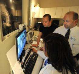
Researchers Jay Hegde and Xing Chen are using functional MRI to look at the brains of study participants learning how to break camouflage in order to help identify soldiers who will be good at it and identify better ways to teach it. Credit: Phil Jones, GHSU Photographer
Researchers want to help the Army better camouflage its soldiers and break the enemy's efforts to hide.
"We want to make our camouflage unbreakable and we want to break the camouflage of the enemy," said Dr. Jay Hegde, neuroscientist in the Medical College of Georgia at Georgia Health Sciences University.
Hegde and GHSU Postdoctoral Fellow Dr. Xing Chen are using a relatively simple technique they developed to teach civilian volunteers to break camouflage. They flash a series of camouflage pictures on a computer screen, providing about a half second after each to spot, for instance, a face in a sea of mushrooms. A green light signals a correct answer and a red light signals an incorrect answer. The computer-generated images include distractions to make the difficult task even more challenging.
They are finding that an hour of daily training in as little as two weeks results in proficiency for 60 percent of the mostly college and graduate school students who have signed up for their training. The Army's current approach is taking soldiers into battlefield situations to hone these skills.
As part of a three-year grant from the Office of Army Research, the researchers want to determine which parts of the brain light up when trained snipers break camouflage.
"We need to figure out how the expert camouflage-breakers do it," Hegde said. "We want to figure out what parts of the brain are most responsive when people break camouflage and, a related experiment is what part of the brain changes its response when people learn to break camouflage." Their techniques include functional magnetic resonance imaging to measure blood flow activity as an indicator of brain cell activity.
Figuring out which parts of the brain are involved could give the Army and others a better way to identify future first-rate snipers and objectively assess instructional efforts.
"If you are the Dean of the Army Sniper Corps and want to develop top-notch snipers, you don't want to spend a year training them before giving up on half of them," Hegde said. A brain scan could help signal whose relevant areas are well developed and, consequently, have natural skill.
Early evidence points toward two regions of the temporal lobe, found on either side of the brain and known to have a role in speech and vision. A region called the fusiform gyrus – which plays a role in facial recognition and lights up when people become experts at recognizing various objects, such as a particular bird species – may be important in breaking camouflage as well.
Hegde suspects that expertise at breaking camouflage stems from the fusiform gyrus in combination with some other area(s) of the brain. And, because good recognition skills don't typically translate from one area to another, he also suspects that the parts of the brain involved vary with the object of their attention. For example, the ability to easily recognize the make and model of a car doesn't guarantee skill at breaking camouflage and Hegde notes that some military snipers aren't good at game-hunting.
Vision happens when light enters the retina where photoreceptor cells turn it into signals that are interpreted by the brain. "If there is a whole lot of light falling on them, they send a lot of signals, beep, beep, beep," he said in rapid succession. "If there is a little bit of light they fire slowly." The brain connects the dots to form a familiar face or landscape. Camouflage complicates the task – the difference between recognizing a mountain goat against a clear blue sky and finding a moth among a pile of fall leaves.
"Here is the beautiful thing that we are finding out: if you know what you are looking for, the next time you can break the camouflage of the moth. Without knowing what you are looking for, the picture also is ambiguous," Hegde said.
Provided by Georgia Health Sciences University


