Tension on gut muscles induces cell invasion in zebrafish intestine, mimicking cancer metastasis

The stiffness of breast tissue is increasingly recognized as an important factor explaining the onset of breast cancer. Stiffening induces molecular changes that promote cancerous behavior in cells. Bioengineering studies have found that breast cancer cells grown on a 3-D gel have enhanced cell replication and decreased organization as rigidity increases. These signals are probably coordinated by surface proteins that communicate with connective tissue, to regulate cell replication, death, and movement. However, very little is known about how stiffness or other physical characteristics of tissues contributes to cancer behavior in living animals.
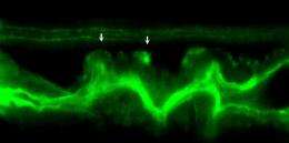
Towards a better understanding of how tissue stiffness drives cancer, in a new paper published in PLOS Biology this week, Michael Pack, MD, associate professor of Medicine, Perelman School of Medicine, University of Pennsylvania, and colleagues, show that epithelial cells lining the intestine of zebrafish that carry an activating mutation of the smooth muscle myosin gene form protrusions called invadopodia that allow the cells to invade surrounding connective tissue. The epithelial cell protrusions form in response to non-regulated contractions in the surrounding smooth muscle cells. These contractions generate oxidative stress in the epithelial cells, which has been linked to invadopodia formation in human cancer cells.
The zebrafish mutant is a novel living model to study how physical characteristics of a tissue can induce cells to invade surrounding tissues, the first step in cancer metastasis.
Epithelial cells of the digestive tract and many other organs are separated from their underlying connective tissue cells by a thin layer of extracellular matrix called the basement membrane. During cell invasion, epithelial cells breach the basement membrane and invade the adjacent connective tissue where the organ's blood vessels and lymphatic channels are located. Invasive cancer cells that are able to enter the blood vessels or lymphatics can migrate to distant tissues thus forming satellite tumors known as metastases.
An important question in cancer research is how invasive cells in living animals correctly localize the enzymes that degrade basement membrane proteins. Investigators have known for some time that many types of cancer cells accomplish this by forming specialized protrusions. It is not known, however, whether these invadopodia are required for cell invasion in living animals or what triggers their formation.
Researchers don't even really know if invadopodia have a normal purpose in healthy tissue. Pack explains that their study was the first to show that invadopodia occur in living animals. This work in live zebrafish is the first direct evidence that invadopodia play a role in tissue cell invasion and identifies the physical signal that drives this process, which in turn, does so by inducing oxidative stress in the invasive cells.
Indeed, eliminating Tks5, a protein required for invadopodia formation in mammalian cells that helps generate reactive oxygen species, blocked formation of the protrusions and rescued mutant zebrafish from cell invasion.
Mouse studies along the same research lines as the zebrafish are planned. The zebrafish and mouse models under development could one day be used to screen potential drugs to stop formation of the errant invadopodia, thus preventing metastases at an early stage of their development. Supporting this idea, mutations in the human smooth muscle myosin genes are present in patients with colorectal cancer and are associated with cancer invasion.
In addition, the team's experiments also point to the possibility that humans who carry a single copy of smooth muscle mutations similar to the zebrafish mutation might inherit a disposition to cancer progression, if they develop a cancer independently of the mutation. The fish with one copy of the mutant gene develop normally but show cell invasion when exposed to drugs that cause oxidative stress, and the risk is greater in fish carrying oncogenes. "This is an interesting finding because oxidative stress is common in cancers and inflammatory conditions that predispose to cancer, such as ulcerative colitis," notes Pack.


