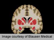
Youth with type 1 diabetes mellitus exhibit a pattern of regional diffusion tensor imaging differences that is suggestive of axonal injury or degeneration and may be related to episodes of severe hypoglycemia, according to a study published online Nov. 8 in Diabetes.
(HealthDay)—Youth with type 1 diabetes mellitus (T1DM) exhibit a pattern of regional diffusion tensor imaging differences that is suggestive of axonal injury or degeneration and may be related to episodes of severe hypoglycemia, according to a study published online Nov. 8 in Diabetes.
Jo Ann V. Antenor-Dorsey, M.P.H., Ph.D., from Washington University in St. Louis, and colleagues used diffusion tensor imaging (region-of-interest and voxelwise tract-based spatial statistics) to quantify white matter integrity in a retrospective study of youth with T1DM and control participants. Medical records and interviews with parents were used to determine exposure to chronic hyperglycemia, severe hyperglycemic episodes, and severe hypoglycemia.
In the T1DM group, the researchers identified lower fractional anistropy in the superior parietal lobule and reduced mean diffusivity in the thalamus. Reduced anisotropy and increased diffusivity in the superior parietal lobule and increased diffusivity in the hippocampus were all associated with a history of three or more severe hyperglycemic episodes.
"These results add microstructural integrity of white matter to the range of structural brain alterations seen in T1DM youth and suggest vulnerability of the superior parietal lobule, hippocampus, and thalamus to glycemic extremes during brain development," the authors write. "Longitudinal analyses will be necessary to determine how these alterations change with age or additional glycemic exposure."
More information:
Abstract
Full Text (subscription or payment may be required)
Journal information: Diabetes
Copyright © 2012 HealthDay. All rights reserved.


