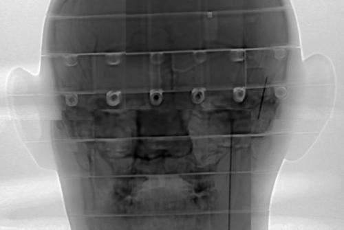
Scientists examine proton radiography of brain mockup

Los Alamos researchers and German collaborators have investigated the application of giga-electron volt (GeV, or billion electron volts) energy proton beams for medical imaging in combination with proton radiation treatment for cancer.
The use of such a high-energy proton beam is ideal for imaging small tumors within patients for targeted proton therapy. Proton radiography, which was invented at Los Alamos, employs a high-energy proton beam to image the properties and behavior of materials.
Significance of the research
Proton beams for cancer treatment use the dramatic increase in energy deposition as the proton stops in tissue to kill the tumors. The incident proton beam must have relatively low energy (approximately 200 MeV) to stop within the human body. However, the low energy proton beam is susceptible to multiple scattering that spreads the beam at the end of its range, just as the protons reach the tumor. This effect restricts the utility of proton therapy to relatively large tumors. The German researchers have discovered that increasing the energy of the proton beam to a GeV reduces the scattering. The GeV proton beam, called a "proton knife," enables proton therapy to be applied to smaller tumors with greater accuracy. Irradiating the tumor from multiple angles allows uniform distribution of protons throughout its volume.
Locating the position of small tumors very accurately is difficult. Presently, X-ray CAT (computed axial tomography) scan results are collected weeks or months before treatment sessions. The "map" of the internal structure from the CAT scan is used to guide the proton beam to the tumor location within the body. This can be problematic as the tumor position can change within the body between the CAT scan and the treatment. Treating smaller tumors requires locating them precisely during the treatment session. A 1 GeV proton beam, which is used for the treatment, could image the tumor within the patient.
Research achievements
The Los Alamos researchers and collaborators investigated GeV proton radiography at LANSCE using biological samples and a high-fidelity human head mockup, called a Matroshka. This mockup was designed and built for dose measurements of humans in the International Space Station. The Matroshka's density and internal structure accurately matches a human head. The team radiographed the mockup with protons to determine the ability of GeV radiography to locate internal features. The scientists observed sub-mm resolution of features and contrast for structures located in soft tissue.


