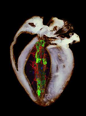
A mouse heart, in gray, shows signs of heart failure because it is missing Mfn2, newly identified as a key molecule in the process that culls unhealthy mitochondria from cells. Superimposed on the mouse heart is a fruit fly heart tube, shown in color. It also shows signs of failure because it is missing Parkin, another key molecule in mitochondrial quality control. These same molecules implicated in heart failure also play roles in Parkinson's disease. Credit: Gerald W. Dorn II, MD
Researchers at Washington University School of Medicine in St. Louis have described a missing link in understanding how damage to the body's cellular power plants leads to Parkinson's disease and, perhaps surprisingly, to some forms of heart failure.
These cellular power plants are called mitochondria. They manufacture the energy the cell requires to perform its many duties. And while heart and brain tissue may seem entirely different in form and function, one vital characteristic they share is a massive need for fuel.
Working in mouse and fruit fly hearts, the researchers found that a protein known as mitofusin 2 (Mfn2) is the long-sought missing link in the chain of events that control mitochondrial quality.
The findings are reported April 26 in the journal Science.
The new discovery in heart cells provides some explanation for the long known epidemiologic link between Parkinson's disease and heart failure.
"If you have Parkinson's disease, you have a more than two-fold increased risk of developing heart failure and a 50 percent higher risk of dying from heart failure," says senior author Gerald W. Dorn II, MD, the Philip and Sima K. Needleman Professor of Medicine. "This suggested they are somehow related, and now we have identified a fundamental mechanism that links the two."
Heart muscle cells and neurons in the brain have huge numbers of mitochondria that must be tightly monitored. If bad mitochondria are allowed to build up, not only do they stop making fuel, they begin consuming it and produce molecules that damage the cell. This damage eventually can lead to Parkinson's or heart failure, depending on the organ affected. Most of the time, quality-control systems in a healthy cell make sure damaged or dysfunctional mitochondria are identified and removed.
Over the past 15 years, scientists have described much of this quality-control system. Both the beginning and end of the chain of events are well understood. And since 2006, scientists have been working to identify the mysterious middle section of the chain – the part that allows the internal environment of sick mitochondria to communicate to the rest of the cell that it needs to be destroyed.
The video clip of an echocardiogram shows a normal mouse heart. Credit: Gerald W. Dorn II, MD
"This was a big question," Dorn says. "Scientists would draw the middle part of the chain as a black box. How do these self-destruct signals inside the mitochondria communicate with proteins far away in the surrounding cell that orchestrate the actual destruction?"
"To my knowledge, no one has connected an Mfn2 mutation to Parkinson's disease," Dorn says. "And until recently, I don't think anybody would have looked. This isn't what Mfn2 is supposed to do."
The video clip of an echocardiogram shows a mouse heart with signs of heart failure because it is missing Mfn2. The same molecules implicated in heart failure also play roles in Parkinson's disease. Credit: Gerald W. Dorn II, MD
Mitofusin 2 is known for its role in fusing mitochondria together, so they might exchange mitochondrial DNA in a primitive form of sexual reproduction.
"Mitofusins look like little Velcro loops," Dorn says. "They help fuse together the outer membranes of mitochondria. Mitofusins 1 and 2 do pretty much the same thing in terms of mitochondrial fusion. What we have done is describe an entirely new function for Mfn2."
The mitochondrial quality-control system begins with what Dorn calls a "dead man's switch."
"If the mitochondria are alive, they have to do work to keep the switch depressed to prevent their own self-destruction," Dorn says.
Specifically, mitochondria work to import a molecule called PINK. Then they work to destroy it. When mitochondria get sick, they can't destroy PINK and its levels begin to rise. Then comes the missing link that Dorn and his colleague Yun Chen, PhD, senior scientist, identified. Once PINK levels get high enough, they make a chemical change to Mfn2, which sits on the surface of mitochondria. This chemical change is called phosphorylation. Phosphorylated Mfn2 on the surface of the mitochondria can then bind with a molecule called Parkin that floats around in the surrounding cell.
Once Parkin binds to Mfn2 on sick mitochondria, Parkin labels the mitochondria for destruction. The labels then attract special compartments in the cell that "eat" and destroy the sick mitochondria. As long as all links in the quality-control system work properly, the cells' damaged power plants are removed, clearing the way for healthy ones.
"But if you have a mutation in PINK, you get Parkinson's disease," Dorn says. "And if you have a mutation in Parkin, you get Parkinson's disease. About 10 percent of Parkinson's disease is attributed to these or other mutations that have been identified."
According to Dorn, the discovery of Mfn2's relationship to PINK and Parkin opens the doors to a new genetic form of Parkinson's disease. And it may help improve diagnosis for both Parkinson's disease and heart failure.
"I think researchers will look closely at inherited Parkinson's cases that are not explained by known mutations," Dorn says. "They will look for loss of function mutations in Mfn2, and I think they are likely to find some."
Similarly, as a cardiologist, Dorn and his colleagues already have detected mutations in Mfn2 that appear to explain certain familial forms of heart failure, the gradual deterioration of heart muscle that impairs blood flow to the body. He speculates that looking for mutations in PINK and Parkin might be worthwhile in heart failure as well.
"In this case, the heart has informed us about Parkinson's disease, but we may have also described a Parkinson's disease analogy in the heart," he says. "This entire process of mitochondrial quality control is a relatively small field for heart specialists, but interest is growing."
More information: Chen Y, Dorn GW. PINK1-phosphorylated mitofusin 2 is a Parkin receptor for culling damaged mitochondria. Science. April 26, 2013.
Journal information: Science
Provided by Washington University School of Medicine


