December 11, 2013 report
Repairing mitochondria in neurodegenerative disease

(Medical Xpress)—The relationship between fine-scale structure and function in the brain is perhaps best explored today by the study of neurodegenerative disease. Disorders like Rett syndrome may be considered developmental in origin—and defined by exotic mechanisms including X-linked inactivation, DNA methylation, and genomic imprinting—but even here, its larger physical pathology evolves through the course of life and continues to be revealed in almost any place that researchers look. When diseases directly involve inputs to the brain like vitamin or diet, and can also be controlled by them, things get even more interesting. More often than not, these disorders have a clear genetic component, are frequently linked to the mitochondria, and lead to progressive and often perplexing deficits of movement. One such enigma is known as pantothenate kinase-associated neurodegeneration, or PKAN syndrome, in its the most frequent form. A recent open paper in the journal Brain explains.
This particular syndrome can be caused by any number of a hundred or so mutations in the PANK2 gene, which codes for the mitochondrial enzyme pantothenate kinase 2. Of the four nuclear-coded PANK genes, only PANK2 is targeted to the mitochondria. Its protein product is involved in co-enzyme A biosynthesis and catalyzes the phosphorylation of pantothenate (vitamin B5). The hallmark pathology, as defined by T2-weighted MRI, can be seen in the globus pallidus and even has its own unique name— the Eye-of-the-Tiger sign.
The researchers used a mouse model of the disease with a Pank2 double gene knockout. On a standard diet, the mice showed growth issues, azoospermia (lack of sperm) and minor mitochondrial dysfunction, but not some of the other typical issues like iron accumulation in the brain or retinal degeneration. Since co-enzyme A is crucial to several metabolic pathways, the researchers also tested the mice on a high fat ketogenic diet. Under these conditions, ketone bodies produced through fatty acid oxidation bypass the normal glycolytic pathways and proceed directly to the citric acid acid.
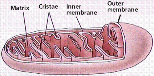
On the ketogenic diet, the mitochondria, which were already ailing with abnormal, swollen cristae, fared much worse, losing some cristae entirely. Extensive lipofuschin deposits were also found in these mice, and movement issues were amplified. It had previously been established in other organisms like flies, that panthethine (a dimeric form of vitamin B5 linked by cysteamine bridging groups) could counteract these issues. When the mice were given panthethine, the general pathology was resolved. In particular, the mitochondria were completely rescued, presumably restored to health, or otherwise replaced in the natural course of events.
The researchers also evaluated mitochondrial membrane potential using dye staining methods. In the knockout mice, membrane potential was compromised, however it was completely restored by the panthethine. At present there is no definitive way to predict functional variables, like membrane potential, from the morphology as it is seen on processed EM tissue. In a recent review of new brain mapping techniques, we discussed this issue, and also pointed to new technologies which may permit closer examinations.
On EM images, one of the most striking features in the interior of mitochondria is the crista junction. This protein structure functionally divides the inner and intermembrane spaces, and controls exchanges between them. While mitochondria come in a variety of forms, the junctions generally converge on a preferred shape and size. Efforts to thermodynamically characterize them in terms of shape entropy have been initiated, as have conceptions of how they evolve as conditions in the mitochondria change mechanically. The so-called "baffle model" of mitochondrial has been entirely replaced by the new cristae junction model which aims to relate structure to function for these organelles, just as we seek it on larger scales for the brain.
Several issues in PNAK style neurodegeneration still stand out like a sore thumb. The iron accumulation is still unexplained, but may be related to another unexplained issue: namely, not only does panthethine fail to cross the BBB, it does not even appear to be working through a vitamin B5 function. When panthethine is metabolized into two pantothenic acid molecules, it also forms two cysteamines. While cysteamine is associated with various side effects, and it can bind and inactivate certain liver enzymes, it also can cross the BBB, perhaps as seen here, to great effect.
The doses necessary for vitamin B5 function are far below those needed here for restorative function. More work is needed to constrain the range of possible mechanisms at play here, but in addition to finding cures for the disease, it will also help cure our ignorance as far as structure-function relations.
More information: Pantethine treatment is effective in recovering the disease phenotype induced by ketogenic diet in a pantothenate kinase-associated neurodegeneration mouse model, Brain (2013) DOI: 10.1093/brain/awt325
Abstract
Pantothenate kinase-associated neurodegeneration, caused by mutations in the PANK2 gene, is an autosomal recessive disorder characterized by dystonia, dysarthria, rigidity, pigmentary retinal degeneration and brain iron accumulation. PANK2 encodes the mitochondrial enzyme pantothenate kinase type 2, responsible for the phosphorylation of pantothenate or vitamin B5 in the biosynthesis of co-enzyme A. A Pank2 knockout (Pank2−/−) mouse model did not recapitulate the human disease but showed azoospermia and mitochondrial dysfunctions. We challenged this mouse model with a low glucose and high lipid content diet (ketogenic diet) to stimulate lipid use by mitochondrial beta-oxidation. In the presence of a shortage of co-enzyme A, this diet could evoke a general impairment of bioenergetic metabolism. Only Pank2−/− mice fed with a ketogenic diet developed a pantothenate kinase-associated neurodegeneration-like syndrome characterized by severe motor dysfunction, neurodegeneration and severely altered mitochondria in the central and peripheral nervous systems. These mice also showed structural alteration of muscle morphology, which was comparable with that observed in a patient with pantothenate kinase-associated neurodegeneration. We here demonstrate that pantethine administration can prevent the onset of the neuromuscular phenotype in mice suggesting the possibility of experimental treatment in patients with pantothenate kinase-associated neurodegeneration.
© 2013 Medical Xpress


