New breast cancer imaging method promising
The new PAMmography method for imaging breast cancer developed by the University of Twente's MIRA research institute and the Medisch Spectrum Twente hospital appears to be a promising new method that could improve on existing imaging technology like X-ray mammography and MRI. Such is the conclusion reached by Michelle Heijblom after well over four years of doctoral research on this method, research that will see her obtain her doctoral degree on 23 April. For her doctoral research, Heijblom conducted clinical studies of patients with suspect abnormalities.
University of Twente researchers had previously developed a new method for detecting breast cancer: PAMmography. For her doctoral research, Michelle Heijblom conducted clinical studies of patients with suspect abnormalities, comparing the effectiveness of the PAMmography method with methods like X-ray mammography, the golden standard in breast cancer examination. Her research has led her to conclude that PAMmography is effective and could improve on the detection methods currently used. "We conducted PAMmography measurements on 73 breast cancer patients and were able to detect the malignancy in nearly all of them."
One important advantage of this new method, Heijblom concludes, is that breast tissue density has no impact on the visibility of the abnormality. This means that, on paper at least, the method can also be used for women below fifty. X-ray mammography is less suitable for detecting breast cancer in these women, as their breast tissue is denser.
Technology
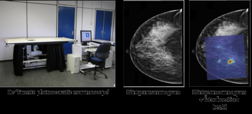
The PAM (Photoacoustic Mammoscope) irradiates the breast with short pulses of light that produce an ultrasound wherever there are large concentrations of blood - like around malignant tumours. The ultrasound next travels from the tumour to the surface skin, where it can be read. During her doctoral research, Heijblom has proven that this new technique is able to detect almost all malignant tumours and also to produce clear images where X-ray images show next to nothing.
Heijblom in her research made used of the first PAM prototype, which employs one light frequency to conduct its measurements. "This was a relatively simple machine, yet still allowed us to see quite a lot." The second, recently developed, prototype is more advanced and measures by two light frequencies. Heijblom believes this will greatly improve the machine's accuracy. Yet she warns against being overly optimistic. "This is entirely new technology. A lot of research and development is required to further improve the method. So it will take years at least before the method can be used in regular healthcare. Still, prospects are positive."
Research
Michelle Heijblom conducted her doctoral research within the Biomedical Photonic Imaging department of the University of Twente's MIRA research institute. Mr Srirang Manohar, PhD, acted as her direct supervisor and Professor Wiendelt Steenbergen, PhD, and Professor Tom van Leeuwen, PhD, were her doctoral thesis supervisors. The research was partially funded by the Netherlands Enterprise Agency (previously known as Agentschap NL). The research was conducted in close cooperation with the Medisch Centrum Twente hospital.


