Imaging gives clearer picture of cancer drugs' chances of success

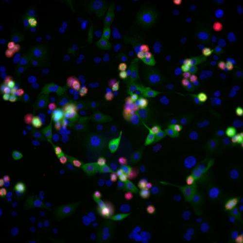
The quest for new cancer treatments could be revolutionised by advances in technology that can visualise living cells and tissues, scientists claim.
Leading edge imaging techniques will make it easier to identify which are the most promising new drugs to take forward for patient testing, a review of the technology suggests.
Applying such techniques early in the drug discovery process could improve the success rate of new medicines by helping to rule out drugs that are unlikely to work.
Researchers at the University of Edinburgh are leading the way in using biological imaging to make the development of new cancer treatments more efficient.
Recent advances mean that scientists can now check how experimental drugs are working inside living cells and in real time.
Using automated microscopes to track fluorescent dyes, researchers can rapidly test thousands of potential drugs in different cancer cell types to find the most promising new treatments.
This pioneering approach – known as phenotypic drug discovery – monitors the effect of a trial drug on the disease as a whole rather than its impact on an individual target protein, which has been the approach until now.
Writing in the journal Nature Reviews Cancer, scientists argue that the new technologies will help to better predict how a drug will work in real life, not just in the test tube.
Currently, just five per cent of drugs tested in clinical trials are approved as therapies for patients. Often drugs do not work in a person as they do in an artificial environment. Sometimes treatments have unexpected side effects that are only picked up in the later stages of the development process.
Report author Dr Neil Carragher, of the Edinburgh Cancer Research UK Centre at the University of Edinburgh, said: "The drug discovery process is hugely expensive and inefficient. In Edinburgh we are leading the way in using biological imaging to streamline the process, allowing us to better select drug candidates with the lowest risk of side effects and the best chances of success in treating patients."
More information: J.R.W. Conway, N.O. Carragher, P. Timpson. Developments in preclinical cancer imaging: innovating the discovery of therapeutics. Nature Reviews Cancer, 17 April 2014. DOI: 10.1038/nrc3724


