(Medical Xpress)—A team of researchers in Germany has created a very highly detailed 3D computer model of an individual rat synapse showing the distribution of approximately 30,000 proteins involved in the process of sending a message from one neuron to another. In their paper published in the journal Science, the team describes how they combined several imaging techniques to create the model, and what it is able to display.
In simple terms, neurons transmit messages between one another via synapses—parts of neurons dedicated to converting electrical signals to chemical signals and vice versa. Synapses generally come in two varieties, the kind that send and the kind that receive signals. Neurons have both kinds of course, which allows them to send and receive messages. To send a message, electrical signals from the neuron travel to the sending synapse where they encounter vesicles. Prodded by the electrical signal, the vesicle releases chemicals called neurotransmitters into the space between (the cleft) the sending synapse on one neuron and the receiving synapse on another. The receiving synapse then processes the message and responds based on the signal it receives.
In the sending synapse, the vesicles are made ready for action by being loaded with neurotransmitters and locked into position. Once they release their cargo, they are moved back out of the way so other vesicles can work while they are being refilled—once ready, they are again placed into position and put to use. This constant recycling goes on over and over, allowing neurons to process a steady stream of messages. In this new effort, the team in Germany has used a variety of microscopic methods to capture the structure of the sending synapse and what happens to it as recycling occurs and messages are sent.
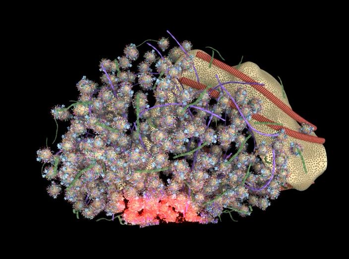
Researchers have designed a three-dimensional model of an “average” synapse. This film shows various views of synapse organization, highlighting components such as the plasma membrane, cytosolic proteins, microtubles, actin, and septin. The camera later zooms in to show the synaptic vesicle and other features at higher resolution. Credit: Wilhelm et al. 2014, Science
To create the model, the researchers isolated rat brain neurons and used mass spectrometry, electron microscopy, super-resolution fluorescence microscopy and antibody staining to get different looks at the sending synapse. In so doing they were able to determine the number of 62 different proteins involved in the recycling process and where they belong in the synapse. That allowed them to build a model able to depict how the synapse actually looks during each stage of the process—a feat that will undoubtedly help many other neuroscientists as they seek to better understand how the brain is able to do all the things it does.
More information: Composition of isolated synaptic boutons reveals the amounts of vesicle trafficking proteins, Science 30 May 2014: Vol. 344 no. 6187 pp. 1023-1028, DOI: 10.1126/science.1252884
ABSTRACT
Synaptic vesicle recycling has long served as a model for the general mechanisms of cellular trafficking. We used an integrative approach, combining quantitative immunoblotting and mass spectrometry to determine protein numbers; electron microscopy to measure organelle numbers, sizes, and positions; and super-resolution fluorescence microscopy to localize the proteins. Using these data, we generated a three-dimensional model of an "average" synapse, displaying 300,000 proteins in atomic detail. The copy numbers of proteins involved in the same step of synaptic vesicle recycling correlated closely. In contrast, copy numbers varied over more than three orders of magnitude between steps, from about 150 copies for the endosomal fusion proteins to more than 20,000 for the exocytotic ones.
Journal information: Science
© 2014 Phys.org


