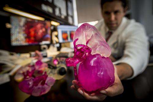
Dr. Jonathan Silberstein holds a replica of a patient’s kidney that was made using a 3D printer. Credit: Paula Burch-Celentano
Most patients rely on their doctors to decipher the black, white and gray images on their CT scans. But what if a patient could instead hold a 3D model made from the CT image in his hands? Suddenly, the picture becomes clearer.
So far, six patients receiving care from the Tulane University Department of Urology have viewed 3D replicas of their kidney cancers prior to surgery.
Ultraviolet lasers are used to print the models in layers using a resin material, similar to plastic. Normal kidney tissue is printed in a clear translucent resin with the tumors in red.
"We can show patients where their tumor is located, how deeply it extends into the kidney tissue, and how we plan to remove it," says Dr. Jonathan Silberstein, chief of the section of urologic oncology.
Until the early 1990s, the standard of care for kidney cancer patients was to remove the entire kidney. Today, many patients have the option of kidney-sparing surgery, through which the tumor is removed, but the healthy portion of the kidney is preserved.
Such organ-sparing surgery may prevent the potential need for dialysis in the future, as well as other issues that could accompany kidney loss.
In addition to the immediate improvement of patient comprehension and assistance in medical trainee education, the models will improve patient outcomes, Silberstein believes.
He says the technology also may be useful for treating other solid-organ cancers, such as liver, lung and prostate cancer. He is applying for a grant to purchase a 3D printer so that the next generation of models can be manufactured in-house.
Provided by Tulane University


