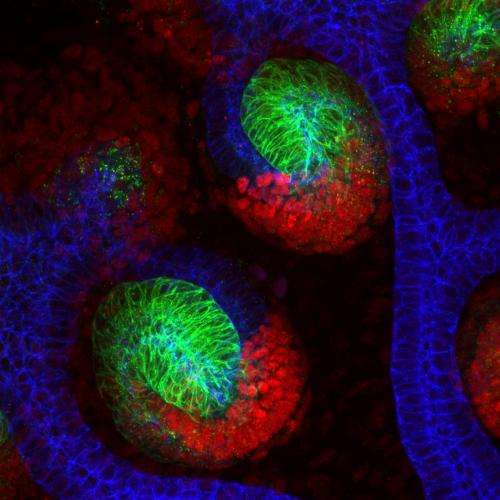
Two nephrons growing in the laboratory. The green and red areas highlight cells that filter waste products from the blood. The blue section shows where urine is collected and taken away for excretion. Scientists have found that a molecule called beta-catenin instructs kidney cells to form these distinct areas of the nephron. Credit: Dr Nils Lindstrom, University of Edinburgh
Striking images reveal new insights into how the kidney develops from a group of cells into a complex organ.
The pictures are helping scientists to understand the early stages of development in mammals.
Researchers at the University of Edinburgh's Roslin Institute used time-lapse imaging to capture mouse kidneys growing in the laboratory on camera.
They identified a key molecule called beta-catenin that instructs cells to form specialised structures within the kidney. These structures - called nephrons -are responsible for filtering waste products from the blood to generate urine.
The images reveal that a gradient in the activity of beta-catenin forms along the growing nephron. It is the concentration of the molecule that instructs cells to form each particular part of the structure.
By changing the activity of beta-catenin in different places, the researchers learned that they could instruct cells to form different parts of the nephron.
If nephrons do not work correctly, it can lead to a wide range of health problems—from abnormal water and salt loss, to dangerously high blood pressure. The findings will help scientists to grow nephrons in the lab that can be used to study how kidneys function.
The use of time-lapsed imaging means that, rather than requiring different litters of mice to study different developmental stages, the same animals can be studied over time. This leads to a significant reduction in the number of animals needed for this type of research.
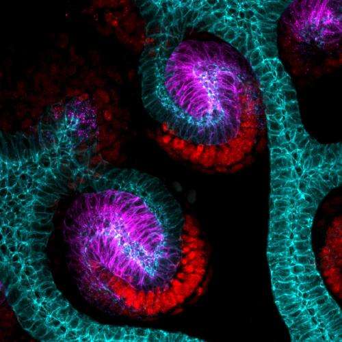
Two nephrons growing in the laboratory. The purple and red areas highlight cells that filter waste products from the blood. The blue section shows where urine is collected and taken away for excretion. Scientists have found that a molecule called beta-catenin instructs kidney cells to form these distinct areas of the nephron. Credit: Dr Nils Lindstrom, University of Edinburgh
Dr Peter Hohenstein, of the University of Edinburgh's Roslin Institute, said: "This is the first time we have been able to identify the molecular signals that instruct cells exactly how to form functional nephrons."
Dr Nils Lindstrom, of the University of Edinburgh, said: "By using time lapse imaging, we can get detailed information about the signals that control how kidneys form at different time-points in development. This means that we can use fewer animals and obtain much more information than normal imaging techniques."
This movie of a mouse kidney growing in the laboratory reveals how a molecule called beta-catenin instructs the organ to develop. The areas in yellow show where the molecule is most active. Credit: Dr Nils Lindstrom, University of Edinburgh
More information: N.O. Lindstrom et al. Integrated β-catenin, BMP, PTEN, and Notch signalling patterns the nephron. eLife, February 2015. elifesciences.org/content/4/e0 … sthash.SE7jmGZa.dpuf
Journal information: eLife
Provided by University of Edinburgh


