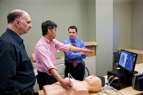
Deep muscle activity exposed via M-mode ultrasounds

M-Mode ultrasound is being tested in a variety of studies as a non-invasive, clinical tool to assess deep muscle activity, as opposed to electromyography which is widely considered to be highly invasive and painful.
The four studies, which are being conducted at Curtin University, explore how M-mode ultrasound can be used to capture the motion of connective tissue within and around muscle.
Lead author Professor Angela Dieterich says M-mode ultrasound is widely used in echocardiography to assess heart motion, allowing doctors to record highly accurate measurements of the motion of deep and superficial muscles.
"Normally, muscle activity is measured using electromyography which records the electrical signals muscles produce while active," Prof Dieterich says.
"Adhesive electrodes are attached on to skin, which creates no discomfort for the subjects, however, only about 10 per cent of the human muscles can be measured by this surface electromyography."
She says to target deeper muscles, needles or hair-fine wires need to be injected into the muscle, which can be uncomfortable, painful, and is not widely used.
"Evidence strongly suggests that the activity of deep muscles is most affected in painful conditions," Prof Dieterich says.
"Our goal was to establish a non-invasive, harmless method for measuring deep muscle activity that would enable painless, risk-free access to many more muscles than is currently possible.
"Such a method would provide information on the unconscious control of movement, with the potential of changing the current physical therapy of painful musculoskeletal conditions."
M-mode ultrasound similar to conventional treatment
Prof Dieterich says the M-mode procedure is the same as conventional ultrasound, and works by a practitioner holding a probe with a bit of gel on the skin.
"The ultrasound beam is harmless with no side effects in clinical applications," she says.
"Motion onset is very obvious in M-mode traces, anyone can see it—it is recognised by an interruption of the straight lines that indicate relaxed muscle.
"We developed a method for computed motion detection to provide reliable, objective measurement points."
However, Prof Dieterich says the method needs to be refined and verified on more muscles.
"We are currently doing this in collaboration between Curtin and three European groups," she says.
"On the other side, the clinical usefulness of M-mode measurements needs to be explored; how do muscles move normally, how does motion change with pathology or with ageing, and how can we address deep muscles by exercises?"
More information: "M-mode ultrasound used to detect the onset of deep muscle activity" DOI: dx.doi.org/10.1016/j.jelekin.2014.12.006


