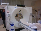
(HealthDay)—Magnetic resonance imaging can be used to visualize the effects of continuous traction on herniated lumbar intervertebral discs and their surrounding structures, according to a study published in the June issue of Radiology.
Tae-Sub Chung, M.D., Ph.D., from the Yonsei University College of Medicine in Seoul, South Korea, and colleagues used real-time magnetic resonance imaging to examine the morphologic changes in herniated lumbar intervertebral discs and surrounding structures during lumbar traction. Forty-eight patients with lumbar disc herniation were treated with continuous lumbar traction. Real-time magnetic resonance imaging was performed before traction initiation and at 10-minute intervals during 30 minutes continuous traction of 30 kg.
The researchers observed changes in disc shape, disc reduction with opening in the intervertebral disc, reduction of herniated disc volume, separation of the disc and adjoining nerve root, and widening of the facet joint as a result of continuous traction on herniated lumbar discs and surrounding structures. There were increases in the mean lumbar vertebral column length (1.45 percent elongation after 30 minutes; P < 0.001) and the mean disc reduction ratio with time of traction (8.57, 15.24, and 17.94 percent after 10, 20, and 30 minutes, respectively).
"The results of this study demonstrated that the real-time effects of continuous traction on herniated lumbar intervertebral discs and their surrounding structures can be visualized by using magnetic resonance imaging," the authors write.
More information: Full Text (subscription or payment may be required)
Journal information: Radiology
Copyright © 2015 HealthDay. All rights reserved.


