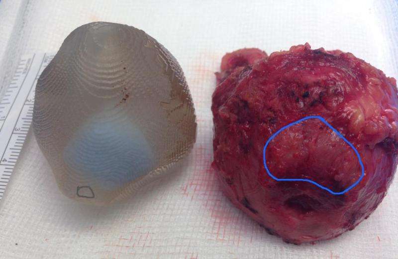
3D printed prostate on the left and the removed specimen on the right showing the chestnut shape with a base wider than the apex, and the location of the tumour which is defining the shape.
At the London Clinic, Professor of Urology Prokar Dasgupta successfully removed a prostate cancer tumour from a patient, 65-year old Mr Alexander Spyrou from London, using an exact replica of the patient's prostate, complete with tumour.
The 3D printed prostate is the brainchild of Dr Clare Allen, consultant radiologist at the London Clinic, and stands to revolutionise the way prostate surgery is carried out, especially when the tumour is in a particularly critical location.
Prof Dasgupta explains: "One of the disadvantages of doing prostate surgery the way I do it, with robotics, is the lack of touch. While you can see things better in 3D, HD, magnified 10x, you lose this crucial sense of touch. In this patient, I could feel the tumour in the 3D model and feel how close the tumour was to the surface. Normally, we plan where the tumour is in our minds but here I held the model in my hand as I performed the procedure with the Da Vinci Robot, where I'm seated remotely at a console. The model allows for better planning and accuracy which is what you want to hear in cancer surgery.
"There is another aspect to it—the nerves for erections are on the sides of the prostate. We have spared these nerves. The cancer in Mr Spyrou has been removed successfully and additionally, he was continent straightaway, with no reduction in his quality of life post-surgery."
As Dr Allen explains: "The tumour in Mr Spyrou's case was in a very difficult location, very close to the muscle which provides continence – the sphincter muscle. The distance between the two was only 1mm and we wanted to remove all the cancer successfully without disturbing the sphincter muscle."
Alexander Spyrou was the first patient to take advantage of the new technology in November 2015. Whilst radiotherapy was an option, Mr Spyrou opted for surgery and when the new technology became available, he was happy to be the first to benefit from it. The operation took about 2 hours and involved 6 small incisions in the stomach area to allow access for the robotic arms. These were closed with staples that were removed after seven days, causing no pain.
He explains: "I naturally felt apprehensive but having involved my wife and discussed the matter at every stage with her, and after meeting with Professor Dasgupta, I felt very comfortable and could not wait to have the procedure done as quickly as possible. As it was at an early stage and there was evidence of cancer I felt it was important to deal with the problem as quickly as possible. There was no point in 'burying my head in the sand'.
"It's only two months since my surgery and my recovery period is ongoing, however, once the catheter was removed after a week at home, I had immediate control of my bladder function, which was wonderful. I am now feeling better every day and looking forward to getting back to a full and active life including travelling and sailing.
"The staff at the London Clinic were kind, discreet and caring, both before and after the procedure and I felt very comfortable about going forward with the surgery there, I would recommend anyone in my situation to go forward for surgery at their earliest possible opportunity."
Mr Mark Feneley, consultant urologist at the London Clinic who referred Mr Spyrou to Professor Dasgupta says, "Using this 3D technology here at the London Clinic perfectly demonstrates just how much a multi-disciplinary team can achieve. From myself at the diagnostic stage, to Dr Allen in Radiology and Professor Dasgupta as the surgeon, it has brought together all avenues of our expertise and is a truly inspiring achievement."
How the 3D prostate was made
From the patient's MRI scan, Dr Allen was able to mark out where the tumour was and then using MIM software, generated the 3D model which was printed by Nuada Medical. Prior to surgery, other surgical assessments and techniques had to be undertaken in order to precisely confirm the diagnosis and the location of the cancerous lesion within the prostate gland. With 3D printing, the surgeon has the virtual prostate in their hand and is able to plan things far more accurately.
After the first meeting three months ago, where the concept was shared with the consultant, Nuada Medical and Dr Allen have worked closely together to develop the software recording process that provides 3D images of the prostate and any abnormal lesions (tumours) within it.
Provided by The London Clinic


