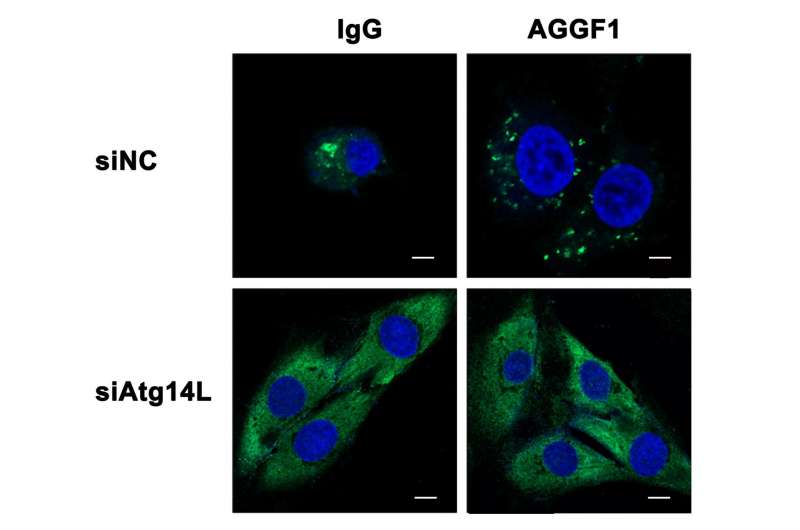
Atg14L siRNA inhibited autophagy induced by AGGF1. The number of DFCP1-positive puncta was increased by AGGF1 treatment in HUVECs. Knockdown of Atg14L using siRNA significantly decreased the number of DFCP1 puncta and blocked the effect of AGGF1. Scale bar = 10 μm. Credit: Lu et al. (2016)
Coronary artery disease, the number one killer world-wide, restricts and ultimately blocks blood vessels, and cuts off oxygen supply to the heart. A study published on August 11 in the open access journal PLOS Biology reports that treatment with AGGF1, a protein which promotes angiogenesis (the growth of new blood vessels), can successfully treat acute heart attacks in mice. The therapeutic benefits depend on autophagy, a normal breakdown process that removes cellular structures that are damaged or no longer needed and recycles their molecular components.
Therapeutic angiogenesis is an experimental treatment that uses the body's own factors to grow new blood vessels, which in turn can serve as endogenous bypasses of blocked older vessels. Qing Kenneth Wang from Huazhong University of Science and Technology in Wuhan, P. R. China and the Cleveland Clinic in Cleveland, USA, and colleagues study the potential of AGGF1, an angiogenesis-promoting protein present in mice and humans, as a therapeutic agent.
Blood vessels are formed by endothelial cells, and the researchers initially examined the effects of AGGF1 on human endothelial cells. One of the earliest responses to AGGF1 exposure, they found, is the induction of autophagy. Autophagy was detected by molecular markers as well as characteristic changes to the cellular morphology. When the researchers looked at the hearts of mice treated with AGGF1, they found that not just endothelial cells but also other cells types in the heart respond with autophagy.
To examine the connection between AGGF1 induction of both autophagy and angiogenesis, the researchers used drugs that inhibit autophagy. Treatment of endothelial cells exposed to AGGF1 with these drugs, they found, blocks blood vessel formation at the earliest stages, suggesting that autophagy is required for AGGF1-mediated angiogenesis.
When the researchers created mice with mutations in either one or both copies of the Aggf1 gene, they found that mice that don't have any AGGF1 die as embryos. About 60% of mice that have one intact copy, however, survive to adulthood, and those mice have reduced levels of autophagy in their hearts.
The researchers then turned to a mouse model of myocardial infarction (MI). They found that AGGF1 levels increase in the injured heart tissue heart after MI, and that this is likely mediated by hypoxia, i.e., the lack of oxygen, in the affected tissue. Treatment with external AGGF1 following MI increases the number of mice that survive two and four weeks later. It also substantially improves parameters measured by echocardiography as well as other cardiac functions in the treated mice.
Looking at the mechanisms, the researchers report that AGGF1 induces autophagy and angiogenesis, increases the number of cardiac cells that survive, and reduces scarring in the heart compared with mice that had an acute MI but did not receive AGGF1. The researchers also identified a key molecular regulator called JNK that mediates the induction of autophagy by AGGF1; drugs blocking JNK prevent the induction of autophagy by AGGF1. And blocking autophagy by drugs or mutations in genes required for the process abolishes the ability of AGGF1 to promote angiogenesis and cardiac repair after MI, demonstrating again an essential role of autophagy in mediating the beneficial effects of AGFF1.
Acknowledging that it is not yet clear how autophagy activates angiogenesis, the researchers conclude that their data nonetheless "uncover new fundamental molecular mechanisms underlying autophagy and therapeutic angiogenesis and provide a novel treatment strategy for [coronary artery disease] and MI, the leading cause of sudden death worldwide".
More information: Lu Q, Yao Y, Hu Z, Hu C, Song Q, Ye J, et al. (2016) Angiogenic Factor AGGF1 Activates Autophagy with an Essential Role in Therapeutic Angiogenesis for Heart Disease. PLoS Biol 14(8): e1002529. DOI: 10.1371/journal.pbio.1002529
Journal information: PLoS Biology
Provided by Public Library of Science


