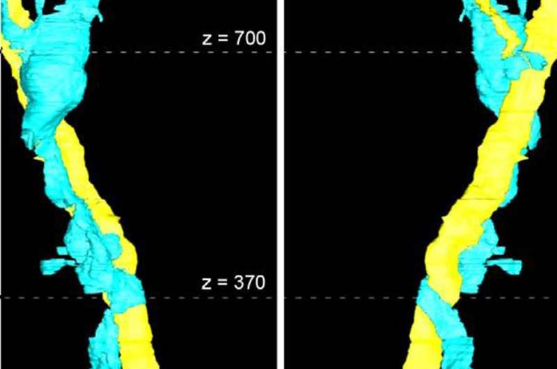
3-D reconstruction of a collagen cable (yellow) and collagen-producing cell (blue). Credit: Dr. Allison Gillies and Dr. Richard Lieber
Eight million people per year in the UK suffer from muscular diseases and injuries including muscular dystrophy, cerebral palsy, exercise-related injuries, rotator cuff tears, and age-related muscle loss.
A new form of 3D imaging of muscles has allowed researchers to "see" inside muscle and trace long cables made up of a protein called collagen. Collagen cables are one culprit behind muscular diseases and injuries, so targeting them could provide treatments.
That's according to new research from a team at UC San Diego and the Rehabilitation Institute of Chicago (RIC) led by Dr. Richard Lieber, currently Chief Scientific Officer at RIC and published in The Journal of Physiology.
Muscular conditions, whether hereditary, exercise-induced, or due to normal aging, can result in stiff, dysfunctional muscles due to changes called fibrosis. Fibrosis is a roadblock to muscle recovery, and can result in muscle pain, weakness, limited range of motion, or require surgery to treat it.
Researchers used a mouse model of skeletal muscle fibrosis to investigate the structure and function of collagen. They visualized collagen with conventional 2D and a newly developed method of 3D electron microscopy, mechanically measured muscle stiffness, and quantified the collagen producing cells.
Dr. Allison Gillies, first author of the study, said:
"The first time we looked at the 3D imaging results we were surprised—the collagen structures that we saw did not fit the textbook definition of muscle."
Collagen had not been previously known to form long chains in muscle. Researchers had seen collagen outside muscle cells, but had not determined the level of organization that could only be visualized by 3D microscopy.
When muscles become fibrotic, the number of cables and cells that produce collagen both increase. These collagen cables and the cells that produce collagen are thus two enticing targets for treating muscle disease and injury.
Commenting on the study, senior author, Dr. Lieber said:
"Reducing the amount of collagen cables or collagen producing cells in fibrotic muscle may improve muscle function and reduce pain, even obviating the need for corrective surgery."
More information: Allison R. Gillies et al, High resolution three-dimensional reconstruction of fibrotic skeletal muscle extracellular matrix, The Journal of Physiology (2016). DOI: 10.1113/JP273376
Journal information: Journal of Physiology
Provided by The Physiological Society


