New way to see artery damage before heart disease sets in

Researchers have developed a new way to non-invasively peer into a person's arteries, detect inflammation, and possibly ward off heart disease before it becomes too severe to treat, a study said Wednesday.
Heart disease is the top killer of men and women in the United States, accounting for one in four deaths nationwide. Each year, about 750,000 Americans have a heart attack.
For decades, doctors have relied on CT scans and angiograms to detect coronary artery disease, a leading cause of heart attack.
These tests focus on finding vessels that are narrowing due to a buildup of cholesterol and other material, called plaque, restricting blood flow to the heart. But they are far from perfect.
Often, patients with narrowed coronary arteries find out only when their condition is severe.
Nor is narrowing of the arteries always a signal of an approaching heart attack.
Instead, inflammation is the real culprit when it comes to triggering a blockage in an artery that leads to heart attack, said researcher Keith Channon, professor of cardiovascular medicine at the University of Oxford.
"Until now, there's been no way to detect inflammation in the coronary arteries," he told reporters on a conference call ahead of the release of the paper in the journal Science Translational Medicine.

"And this is where our new research findings come in."
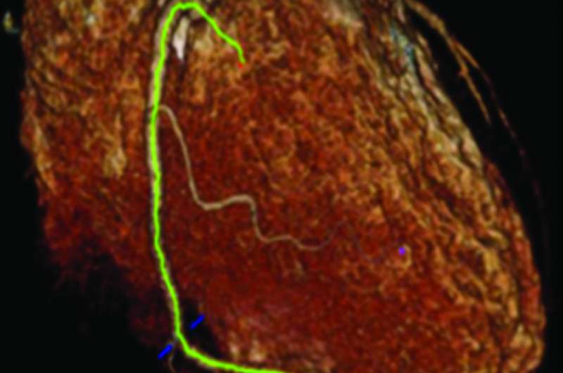
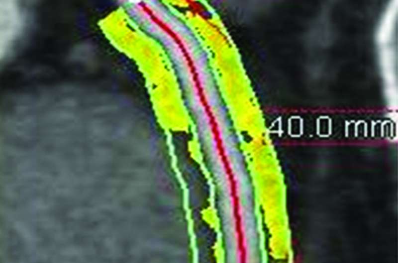
The process works by analyzing changes in the fat tissue that surrounds arteries, known as perivascular fat.
This fat becomes more watery and less fatty when it sits near an inflamed artery, researchers said.
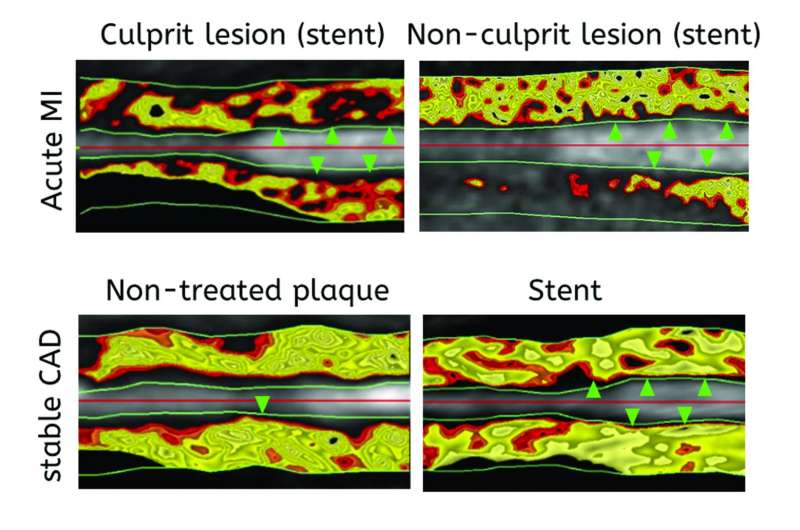
Using a metric called CT fat attenuation index (FAI), researchers have found signs of inflammation in existing CT scans.
This indicator points to either stable coronary atherosclerotic plaques, or ruptured plaques in patients who had recently experienced heart attack.
They also found they could track changes in perivascular fat over time, allowing doctors to detect early signs of disease that might be prevented with interventions like cholesterol-lowering statins.

More study is needed before researchers can say how well the approach may be able to predict future heart attacks and save lives.
"If this is confirmed in larger studies, then that will offer an additional option, an additional readout on standard CT angiography," said Charalambos Antoniades, associate professor of cardiovascular medicine at the University of Oxford.
"I'm optimistic. I believe that, yes it will be able to predict future events. However, we need to wait for the study to be completed."
The results are expected to be published by the end of the year, he said.
More information: "Detecting human coronary inflammation by imaging perivascular fat," Science Translational Medicine (2017). stm.sciencemag.org/lookup/doi/ … scitranslmed.aal2658
© 2017 AFP


