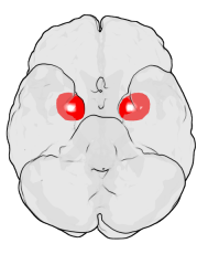
Connections between two brain regions linked with financial risk tolerance

Researchers have known that connections between two areas of the brain, the amygdala and the medial prefrontal cortex (mPFC), have been implicated in the development of affective disorders like depression and anxiety. But new research suggests that this same brain system plays a role in a person's ability to tolerate economic risk.
In a paper published in Neuron on April 5, scientists at the University of Pennsylvania found that structural and functional connections between the amygdala and the mPFC are associated with individual differences in the degree to which a person accepts risk in order to achieve a greater financial return.
The researchers studied 108 healthy young adults. Participants were asked to answer several questions involving their comfort with financial choices—each involving various levels of risk and reward. "We assessed risk tolerance by giving people an opportunity to gamble," explains author Joseph W. Kable. They faced over 120 different scenarios involving the risk of making more or less money. "We assessed how willing individuals were of accepting the risk of getting nothing for the chance of getting a higher amount of money," he said. The study identified individuals ranging from extremely risk-averse all the way to risk-seeking.
Separately, using MRI imaging and diffusion tensor imaging (DTI), the researchers measured the structural and functional connections between various parts of the participants' brains, homing in on the amygdala/mPFC system. They also measured the size of the amygdala, including volume of gray matter and white matter. To make an assessment of an individual's risk tolerance, the scientists correlated their assessment of participants' risk tolerance with the measures of brain structural and functional connectivity.
"The three measurements—structural and functional connections and the volume of amygdala gray matter—reinforce each other to suggest that there is something important about the function of this system related to differences in how tolerant people are to taking risk," adds Kable. "Just by looking at these features of your brain, we could have a reasonable idea if you are someone who will take lots of risk or not."
Specifically, individuals with higher tolerance for risk in the study possessed a larger amygdala (more gray matter) and more functional connections between the amygdala and mPFC as measured by MRI. And higher risk tolerance was identified in individuals with fewer structural connections or pathways between these areas, as measured by DTI.
Going forward, the researchers plan on collaborating with financial planning organizations to see how these brain-system findings can be used as a marker for risk tolerance involving larger economic-based decisions. "Perhaps we can get a better assessment for someone's economic risk tolerance to provide the best advice possible for that particular individual," says Kable. "The idea of using these brain markers and pairing them with some questionnaire or other assessments may help determine a better, more well-rounded sense of your tolerance for risk."
More information: Neuron, Jung et al.: "Amygdala functional and structural connectivity predicts individual risk tolerance" www.cell.com/neuron/fulltext/S0896-6273(18)30196-X , DOI: 10.1016/j.neuron.2018.03.019


