Balance of biomolecular signals stimulates healing by setting skin cells into motion

After a flesh wound, skin cells march forward to close the gap and repair the injury. Findings from a team led by Leah Vardy at A*STAR's Skin Research Institute of Singapore now demonstrate how a carefully regulated set of molecular cues helps coordinate this healing migration.
Vardy was particularly interested in a trio of organic molecules known as polyamines, which play a role in cellular proliferation. "They are well studied in cancer, but much less is known about how changes in their levels can drive normal cellular behavioural changes," says Vardy. An enzyme called AMD1 facilitates the conversion of one polyamine, putrescine, into two other polyamines, spermidine and spermine. As this step is essential in determining the final balance of the three molecules within the cellular environment, Vardy's team examined what happens to AMD1 levels during wound healing.
To model skin damage, they put a scratch on a sheet of cultured human skin cells, and observed a spike in AMD1 levels among cells near the edge of the wound within an hour of the injury. Although initially diffuse, AMD1 expression became more strongly focused within the outermost rows of cells over the next several hours. The researchers then subjected the cultured cells to a gene expression-modulating treatment that greatly reduced their ability to produce AMD1. This resulted in markedly slower healing in the experimental wound model, which the researchers attributed to the imbalance in the relative levels of putrescine, spermidine and spermine. Closer analysis of the treated cells confirmed this, revealing a 2.7-fold increase in putrescine and a two-fold reduction in spermine levels, while spermidine levels remained relatively unchanged.
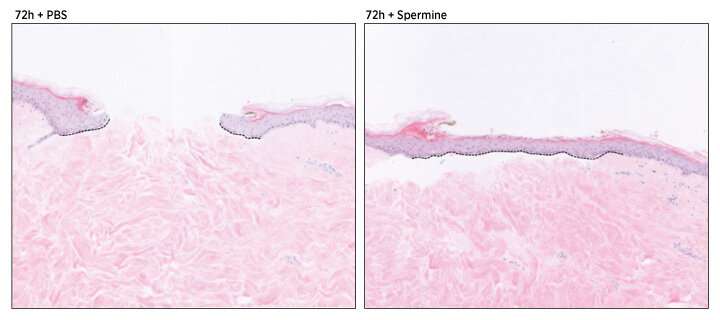
Strikingly, when Vardy and colleagues compensated for this imbalance by adding spermine back to the damaged skin cell cultures, they observed a clear increase in the rate of wound closure. "Low putrescine and high spermine levels are required for cell migration at the wound edge," explains Vardy. They further determined that these polyamines appear to coordinate a reorganization of the cytoskeletal proteins that provide the infrastructure for cell migration during the wound healing process.
Vardy believes that this process may be disrupted in patients whose wounds won't heal after injury, and she is now looking into the potential for polyamine-based therapeutics for wound care. "We are currently addressing the mechanism by which spermine promotes cell migration," she says, "and working with clinicians here in Singapore to determine how polyamine levels are altered in non-healing wounds."
More information: Hui Kheng Lim et al. Polyamine Regulator AMD1 Promotes Cell Migration in Epidermal Wound Healing, Journal of Investigative Dermatology (2018). DOI: 10.1016/j.jid.2018.05.029


