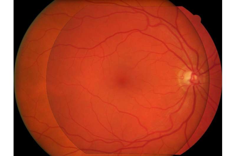
Credit: IPRI
We have introduced an efficient method to superimpose eye fundus images from persons with diabetes screened over many years for diabetic retinopathy. The method is fully automatic and robust to camera changes and colour variations across the images both in space and time. All the stages of the process are designed for longitudinal analysis of cohort public health databases where retinal examinations are made at approximately yearly intervals.
In this paper a method is presented for superimposition (i.e. registration) of eye fundus images from persons with diabetes screened over many years for diabetic retinopathy. The method is fully automatic and robust to camera changes and colour variations across the images both in space and time. All the stages of the process are designed for longitudinal analysis of cohort public health databases where retinal examinations are made at approximately yearly intervals.
The method relies on a model correcting two radial distortions and an affine transformation between pairs of images which is robustly fitted on salient points. Each stage involves linear estimators followed by non-linear optimisation. The model of image warping is also invertible for fast computation. The method has been validated on a simulated montage and on public health databases with 69 patients with high quality images (271 pairs acquired mostly with different types of camera and 268 pairs acquired mostly with the same type of camera) with success rates of 92% and 98% and five patients (20 pairs) with low quality images with a success rate of 100%. Compared to two state-of-the-art methods ours gives better results.
More information: G Noyel et al. Superimposition of eye fundus images for longitudinal analysis from large public health databases, Biomedical Physics & Engineering Express (2017). DOI: 10.1088/2057-1976/aa7d16
Provided by CORDIS


