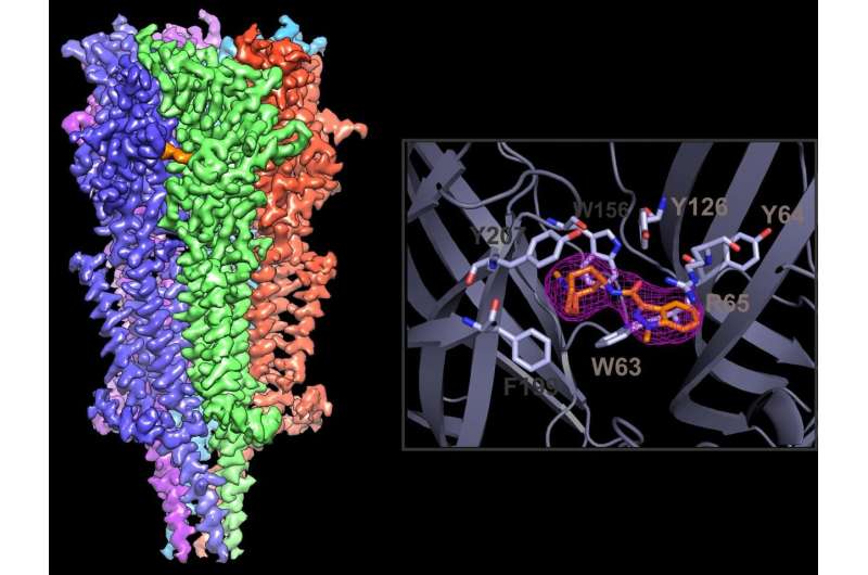
A 2.9 Å cryo-EM reconstruction of the 5-HT3A receptor complex with granisetron. A close-up view of the drug-binding pocket and the density map for granisetron. Credit: Case Western Reserve University School of Medicine
A new study using a special type of electron microscope using samples cooled to extremely cold temperatures provides critical information for drug developers seeking to reduce nausea and vomiting side effects of cancer treatments. Published in Nature Communications, the study offers a glimpse into how widely-used anti-nausea drugs attach to their target protein in the gastrointestinal tract. High-resolution images obtained by this method provide key details about how the drugs attach into a binding pocket on the protein—and offer clues into how their design might be improved.
The study focused on a specific class of drugs used to manage nausea, vomiting, and irritable bowel syndrome, called setrons. Setrons are generally well-tolerated, but some cancer patients do not respond to them, explained study lead Sudha Chakrapani, Ph.D., associate professor of physiology and biophysics at Case Western Reserve University School of Medicine.
"Cancer patients who have vomiting later in their treatment plans—delayed emesis—don't tend to respond to setrons," Chakrapani said. "There is a constant need for better drugs." Drug improvement has been stalled by a lack of models showing exactly how drugs like setrons attach to their target protein in the body—the serotonin (3) receptor. Without a precise model, drug developers have been unable to understand exactly which elements of setron-receptor interactions are most important, and how to enhance them.
The new study provides the highest-resolution images to date of a setron settling inside the binding pocket of a serotonin (3) receptor. Researchers tracked the receptor-drug interactions, to less than a billionth of a meter—using a cryo-electron microscope. Cryo-electron microscopy (cryo-EM) has only recently become available for small protein targets and was the focus of the 2017 Nobel prize in chemistry.
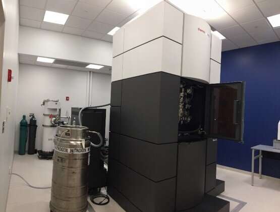
The high-resolution images were collected on a Titan Krios cryo-electron microscope in collaboration with colleagues at Stanford University. The installation of the first Titan Krios microscope at the cryo-EM Core here at Case Western Reserve has just been completed and is now one of two operational microscopes in Northeast Ohio. Credit: Case Western Reserve University School of Medicine
Cryo-EM images revealed setrons use the same attachment site as the receptor's natural binding partner in the body, serotonin, but take a slightly different "pose" that changes the receptor shape slightly. The differences helped the researchers build a more precise model of how setrons work on a molecular level.
Said Sandip Basak, co-first author on the paper, "In the past, we didn't have the confidence to model the drug in its binding pocket. Now we can precisely do that. We can also watch the drug move in the pocket using molecular dynamics simulations."
Chakrapani collaborated with colleagues at Mt. Sinai to identify the most stable interactions between setrons and serotonin receptors. The team watched as setrons twisted and turned in the pocket, revealing key portions of the drug and the receptor that are required for a tight connection. They then mutated the key portions, which eliminated setrons' affinity for the serotonin receptors. Together, the experiments helped reveal which portions of setrons and serotonin receptors are most important, and might be most promising to enhance therapeutically.
"Identifying the binding pocket and the interactions that are most important, and the orientation of the drug in the binding pocket, lays the foundation for designing drugs that are going to be more efficient," said Yvonne Gicheru, who is a co-first author on the paper.
The high-resolution images were collected on a Titan Krios cryo-electron microscope in collaboration with colleagues at Stanford University. The installation of the first Titan Krios microscope at the cryo-EM Core here at Case Western Reserve has just been completed and is now one of two operational microscopes in Northeast Ohio.
More information: Sandip Basak et al, Molecular mechanism of setron-mediated inhibition of full-length 5-HT3A receptor, Nature Communications (2019). DOI: 10.1038/s41467-019-11142-8
Journal information: Nature Communications
Provided by Case Western Reserve University


