Mechanism for the formation of new blood vessels discovered

Researchers from Uppsala University have revealed for the first time a mechanism for how new blood vessels are formed and have shown the importance of this mechanism for embryo survival and organ function. The results could be developed to control the formation of new blood vessels in different diseases. The new study is published in the journal EMBO Reports.
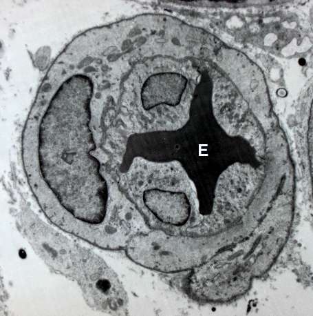
Having a healthy network of blood vessels that bring oxygen and nutrients to tissues is essential for survival. Blood vessels are lined with cells called endothelial cells, which are needed to form the hollow vessel through which blood is channeled to all tissues.
Lena Claesson-Welsh's research group has revealed the mechanism whereby new endothelial cells are formed by the division of existing cells. The loss of this mechanism, resulting in too few blood vessels, may lead to death during embryo development due to an inadequate supply of oxygen to the brain. In other cases, having fewer blood vessels disturbs organ function after birth.
Interestingly, the importance of this mechanism varies with the genetic composition of the individual. In certain genetic designs, the effect is only seen during embryo development, while in other genetic designs, the effect is seen only after birth.
"We were very surprised to discover the importance of genetics in controlling the mechanism of endothelial cell division. Certain types of mice would not survive embryo development, while others would survive but show defects after birth," says Chiara Testini, first author of the study, which was a team project headed by Lena Claesson-Welsh at the Department of Immunology, Genetics and Pathology, Uppsala University.
A protein called vascular endothelial growth factor (VEGF) has long been recognized as a master regulator of endothelial cells. It is known to induce these cells to divide and multiply by affecting activities in the cell nucleus. Exactly how this is controlled has eluded explanation. However, in the present study the researchers demonstrate the involvement of a single amino acid in the receptor protein VEGFR2, which binds VEGF on endothelial cells.
The authors devised experiments using mice that lacked the amino acid in question in the VEGFR2 protein. In these mice, which had everything needed to make new blood vessels expect for the particular amino acid, the cells were no longer able to send signals to the nucleus to cause cell division.
"That such a critical mechanism, formation of new blood vessels, is controlled by a single amino acid was really unexpected and we had to check the results again and again using various techniques. We hope these new findings can be developed to manipulate endothelial cell formation to make more or fewer blood vessels dependent on the need," says Lena Claesson-Welsh, Professor at the Department of Immunology, Genetics and Pathology, Uppsala University, Sweden.
More information: Chiara Testini et al. Myc‐dependent endothelial proliferation is controlled by phosphotyrosine 1212 in VEGF receptor‐2, EMBO reports (2019). DOI: 10.15252/embr.201947845


