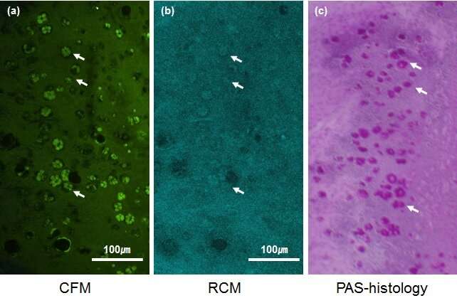
New CGCs fluorescence imaging (a) and the existing CGCs imaging (b,c) are compared. Confocal fluorescence microscopy (CFM) illustrated in (a), confocal reflectivity microscopy (CRM) illustrated in (b), PAS-histology illustrated in (c). The conjunctival goblet cells are marked with white arrows in the figures. Credit: Ki Hean Kim (POSTECH)
Goblet cells are epithelial cells that produce mucins and disperse tears which help the surface of eyes maintain their wet environment. Goblet cells are closely related to autoimmune diseases including dry eyes and chemical burns. Therefore, it is very important to examine the status of goblet cells to better understand and diagnose ocular disease.
Professor Ki Hean Kim and Mr. Seonghan Kim of the POSTECH Department of Mechanical Engineering and Division of Integrative Biosciences and Biotechnology collaborated with Dr. Myoung Joon Kim, who is a former ophthalmology professor at Seoul Asan Medical Center and a doctor at Renew Seoul Eye Center, in finding a better technique of goblet cell assessment. They successfully developed an imaging technology of conjunctival goblet cells with high definition and high image contrast.
The current standard imaging method for goblet cells is impression cytology. This method uses polymer paper attached to the conjunctiva to extract conjunctival goblet cells, and these cells are examined under the microscope after labeling them with special dying agent. However, the method is rarely used because the process is very complicated and it can damage the surface of the conjunctiva.
In the meantime, there has been a report on using confocal reflection microscopy (CRM) to image conjunctival goblet cells. The assessment is non-invasive, yet, it provides relatively low image contrast and it is hard to obtain precise results.
Real-time image of CGCs filmed at 15 frames per second. CFM imaging is shown in left and CRM imaging shown in right. CFM allows real-time imaging of the goblet cells with higher image contrast than the existing imaging methods. Credit: Ki Hean Kim (POSTECH)
In an effort to develop a better imaging method, the research team has studied a clinically compatible cell image technique using fluoroquinolone antibiotics, which are used in eye drops, as fluorescent cell labeling agents. As a result, moxifloxacin, one of the fluoroquinolone antibodies used in eye drops, displayed stronger fluorescence in the goblet cells that are dispersed on the surface of the conjunctiva.
The moxifloxacin-based fluorescence imaging of conjunctival goblet cells was tested in a mouse model. Moxifloxacin was injected into a mouse and it was examined under the CRM after one to two minutes of injection. The team verified that a bright cluster of conjunctival goblet cells on the surface of the conjunctiva was shown. In comparison with the existing CRM, this fluorescence imaging method demonstrated high image contrast and the capacity for real-time imaging at 10 frames per second.
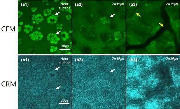
CFM and CRM images of goblet cells from the surface of conjunctiva from a normal healthy mouse. CFM images in figure (a), CRM images in figure (b). Goblet cells are marked with white arrows, blood vessels are marked with yellow arrows. Credit: Ki Hean Kim (POSTECH)
This newly developed high-contrast conjunctival goblet cell imaging is the first of its kind as it uses antibiotics safely applied in ophthalmology nowadays. It can thus be used to develop diagnostic medical devices. Furthermore, it can be utilized in precision diagnosis of dry eye syndromes and evaluation of treatment effect.
Professor Ki Hean Kim explained his future plans, "This study is meaningful in that it overcame the limitation of the conventional imaging method of conjunctival goblet cells. It creatively introduced a non-invasive and high-definition imaging method. We will further advance this imaging method to an assessment of conjunctival goblet cells. We hope to invent a medical device using this method which then can be applied to precision diagnoses of dry eye syndrome and their treatment effect evaluation."
More information: Seonghan Kim et al, In vivo fluorescence imaging of conjunctival goblet cells, Scientific Reports (2019). DOI: 10.1038/s41598-019-51893-4
Journal information: Scientific Reports
Provided by Pohang University of Science & Technology (POSTECH)


