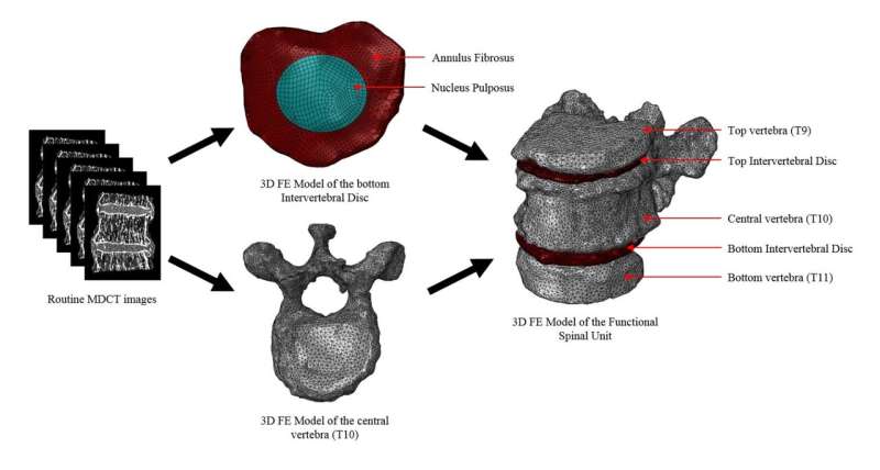
Schematic workflow illustrating methodology from acquisition of images to finite element (FE) modelling of the functional spinal units (FSUs). Credit: SUTD
Osteoporotic vertebral fractures (OVFs) are a prevalent skeletal condition in the elderly, occurring due to a net loss in bone density with the inevitable onset of aging. Unfortunately, they are largely under-diagnosed until detected by clinicians through radiological scans. These fractures have a huge impact on daily lifestyle as the spine is responsible for bodily movements and stability. However, the mechanism behind these fractures remain unclear due to the complex physiological interplay between spinal segments. As such, the fractures are asymptomatic and clinically overlooked until reported by patients.
Extensive research is ongoing in the investigation of alternative biomarkers to assess bone strength and consequently allow prediction of fractures before they are sustained. One such biomarker is the computational prediction of failure load through numerical simulation, also known popularly as finite element analysis. With this analysis, not only is non-invasive examination of the spine possible, but a holistic quantitative evaluation of the bone strength too.
Yet most of the research is focused on the biomechanical analysis of vertebral segments in isolation. The spine consists of many spinal segments, with majority of the load borne by the vertebra and intervertebral discs. Hence, it is essential to include these load-bearing segments when considering the structural strength of spine. Functional spinal units have the advantage of mimicking the biomechanical requirements of the spine better than isolated vertebral segments.
This investigation, recently published in the Spine Journal by the Singapore University of Technology and Design's (SUTD) Medical Engineering and Design (MED) Laboratory, in collaboration with the Technical University of Munich, introduced a semi-automatic computational clinical tool that aims to extract structural information such as failure load from radiological scans of patients using functional spinal units. This study demonstrated that routine clinical scans can be a feasible resource for accurate prediction of OVFs using patient-specific finite element analysis of functional spinal units. These results pave the way for a spinal risk assessment tool to be developed and used by clinicians.
Improved management of OVFs is essential amidst current clinical challenges. Understanding in detail the causes of OVFs will help organizations looking to tackle the increasing morbidity and mortality rates of the aging population, which poses unnecessary socioeconomic burden on society. Implementation of a semi-automatic clinical tool vertebral strength assessment tool could also result in more accurate prediction of osteoporotic fracture risk and aid clinicians with better targeted early treatment strategies.
"Considering the world is aging rapidly, osteoporotic bone fractures are also increasing significantly. So, there is an urgent need to implement computational biomechanical analysis in the clinical scenario since it is a powerful tool for non-invasive evaluation of bone strength. Accordingly, this work lays the foundation for extracting valuable structural information from improved spine models, such as functional spinal units, in the diagnosis of osteoporosis and prediction of OVFs," said lead researcher, Assistant Professor Subburaj Karupppasamy from SUTD's Engineering Product Development pillar.
More information: D. Praveen Anitha et al, Effect of the intervertebral disc on vertebral bone strength prediction: a Finite-Element Study, The Spine Journal (2019). DOI: 10.1016/j.spinee.2019.11.015
Journal information: Spine Journal
Provided by Singapore University of Technology and Design


