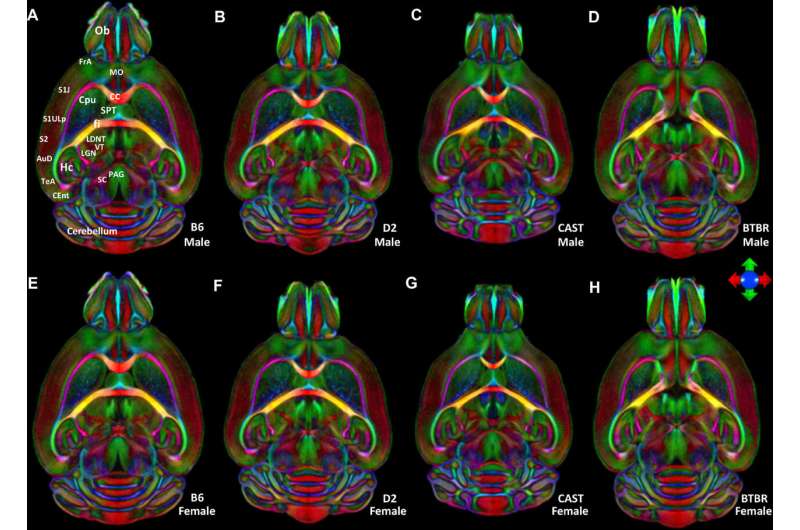
These colorful orbs are maps of the circuitry of mouse brains, showing with unprecedented detail how different areas of the brain are connected. The images were made at Duke's Center for In Vivo Microscopy with magnetic resonance about 90,000 times higher resolution than is used in humans.
In a proof-of-concept study, the technique was found to be even more sensitive at identifying differences in brain structure than the researchers had expected, giving it the potential to study aging, drug exposure and traumatic brain injury.
Credit: Duke University
More information: Nian Wang et al. Variability and heritability of mouse brain structure: Microscopic MRI atlases and connectomes for diverse strains, NeuroImage (2020). DOI: 10.1016/j.neuroimage.2020.117274
Journal information: NeuroImage
Provided by Duke University


