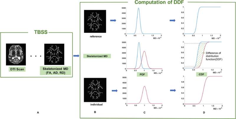
Fig. 1. Flowchart of DDF computation. A: Generating skeletonized MD map (TBSS). B-D: The computation of DDF: Column C shows the PDF for skeletonized MD distribution. Column D shows the CDF for skeletonized MD distribution. The upper row is the reference, the lower row is an individual subject, and row in the middle shows the comparison between individual and reference. The value of DDF is the area with green dash lines. The gray dash lines are the 5th to 95th percentiles of the differences of the inversed distribution functions. Abbreviations: DDF, Difference in distribution functions; TBSS, Tract-Based Spatial Statistics; MD, mean diffusivity; PDF, probability density function; CDF, cumulative distribution function. Credit: DOI: 10.1016/j.neuroimage.2021.118381
Researchers from UNSW Sydney's Centre for Healthy Brain Aging (CHeBA) have developed an improved neuroimaging measure to monitor age-related cognitive decline in older adults.
The findings, published in NeuroImage, indicate that the measure—named "Difference in Distribution Function' will ultimately assist in monitoring the aging process and brain changes seen in Vascular dementias and Alzheimer's disease.
Diffusion Weighted Imaging has been the most widely recognized neuroimaging technique for evaluating the microstructure of white matter in the brain, with white matter integrity critical to normal brain structure and function. Various diffusion weighted imaging measures have been developed to investigate white matter but all have had inherent limitations.
Leader of CHeBA's Neuroimaging Group Associate Professor Wei Wen said that there was a distinct need for an enhanced neuroimaging measure to be developed to overcome these limitations and particularly be able to more effectively and comprehensively describe changes to the white matter.
Lead author Dr. Jing Du said that white matter is important because it is vulnerable to the effect of vascular risk factors.
"It is possible that aging brains suffer significant micro-structural changes due to vascular factors before functional changes are obvious, such as cognitive decline and impact on memory," says Dr. Du.
"The improved measure we have created also differentiates diseased brains from healthy ones," said Dr. Du.
The researchers developed the new measure using study participants from the UK Biobank and validated the measure in healthy brains through CHeBA's Sydney Memory and Aging Study and a study addressing cerebral small vessel disease. The improved diffusion imaging measure has shown to be highly associated with age and cognition across all three cohorts and better explains variance of the changes related to age and cognition than established Diffusion Weighted Imaging measures.
The new measure also has higher sensitivity in distinguishing cerebral small vessel disease patients from cognitively normal participants.
Co-directors of CHeBA and co-authors Professor Perminder Sachdev and Professor Henry Brodaty said that the superior measure shows promise to become a surrogate marker for monitoring the white matter microstructural changes and aging related cognitive decline in older individuals.
"The sensitivity of tracking the brain aging process allows this improved measure to be a biomarker applied for monitoring the structural and functional changes in both healthy and diseased brains, expanding our research potential."
Dr. Du and colleagues are delighted their work is clinically meaningful and immediately useful to the global neuroimaging community, with the computer program for the improved measure already accessible online: bit.ly/CHeBANeuroimaging_DCDF.
More information: Jing Du et al, Difference in distribution functions: A new diffusion weighted imaging metric for estimating white matter integrity, NeuroImage (2021). DOI: 10.1016/j.neuroimage.2021.118381
Journal information: NeuroImage
Provided by CHeBA


