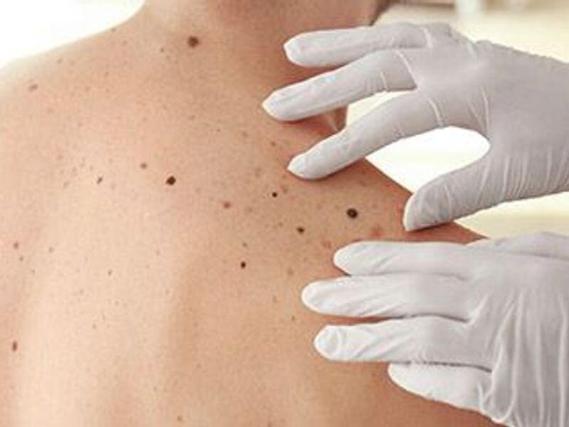
(HealthDay)—Certain dermoscopic structures associated with melanoma detection may have diagnostic importance, including shiny white structures, pseudopods, and blue-white veil, according to a systematic review and meta-analysis published online Aug. 4 in JAMA Dermatology.
Natalie M. Williams, M.D., from the University of Miami Miller School of Medicine, and colleagues examined the diagnostic accuracy of individual dermoscopic structures and patterns used in melanoma detection in a systematic review and meta-analysis. Data were included from 40 studies with 22,796 skin lesions and 5,736 melanomas.
The researchers found that shiny white structures, pseudopods, irregular pigmentation, blue-white veil, and peppering were the structures and patterns with the highest odds ratios for melanoma (odds ratios, 6.7, 6.7, 6.4, 6.3, and 6.3, respectively). Pseudopods, shiny white structures, peppering, and streaks were the structures with the highest specificity (97.3, 93.6, 93.4, and 92.1 percent, respectively), while irregular pigmentation, blue-white veil, atypical network, and a multicomponent pattern had the highest sensitivity (62.3, 60.6, 56.8, and 53.7 percent, respectively).
"The findings of this systematic review and meta-analysis support the diagnostic importance of dermoscopic structures and patterns associated with melanoma detection," the authors write. "The diagnosis of melanoma is usually rendered when the lesion is evaluated as a whole and these features are examined together; thus the more structures that are present in a lesion, the higher the odds the lesion is melanoma."
More information: Abstract/Full Text (subscription or payment may be required)
Journal information: JAMA Dermatology
Copyright © 2021 HealthDay. All rights reserved.


