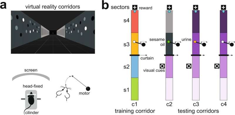
Fig. 1: Behavioral setup: configuration based on virtual environments. a Subjects were head-fixed to metal bars, in turn connected to a stereotaxic frame. Movement was only allowed in a single dimension through a low-friction Styrofoam cylinder placed in front of a curved TFT screen. The movements of the cylinder were recorded by an infrared sensor and converted into virtual reality movements (Arduino Mega board, 2560 Rev3), thus simulating navigation. The self-developed virtual environments were implemented in Unity3D software (Unity Technologies, 2018). b Drawing of the virtual corridors, viewed from above. The virtual corridors were designed with four distinct track patterns on the inner walls, which differed along the longitudinal line. The wall patterns visually define four sectors per corridor, s1–s4, with different profiles between training and test corridors; an intermediate wall (curtain) separates the corridor into two compartments, containing sectors s1 and s2, and s3 and s4, respectively. The walls were designed with colors within the range of wavelengths perceived by the mouse visual system49,50. A final reward zone was identified with a distinctive signal (white cross), where a 1% sucrose drop is provided. In sector 3 of each corridor, a cotton swab attached to a silent motor is slowly advanced to the nose. The swab is moved until it is close enough to the nose to allow contact with the stimuli. In the training corridor at s3, a clean cotton swab is presented, while in the test corridors the cotton is impregnated with different chemosensory cues: a non-pheromonal olfactory cue (sesame seed oil) at c2/s3, male urine at c3/s3 and only a clean cotton swab at c4/s4. Credit: DOI: 10.1038/s41467-021-25442-5
Published recently in Nature Communications, a new study used a model of rodents and connected the influence of feronomic signals on memory for the first time. This will make it possible to explore the deficits of integration of spatial memories and social studies in transgenic models of Alzheimer's disease in the future. The research, directed by professors Enrique Lanuza (Faculty of Biological Sciences) and Vicent Teruel Martí (Faculty of Medicine and Dentistry), has been carried out entirely by research staff from the University of Valencia.
The evidence shows that the generation of the memory of an event or an experience includes various types of information, such as where it happened (spatial component) or who was involved (social component), but how these components are integrated in the brain is still a mystery.
The researchers demonstrated that social and spatial memory are integrated into a circuit formed by three neural structures: the amygdala (part of the brain responsible for emotional reactions), the entorhinal cortex (memory and orientation) and the hippocampus (also related to memory and learning). In the case of rodents, individual recognition depends on pheromonal signals, and this is the first study that shows to what extent this type of information influences memory.
According to Vicent Teruel and Enrique Lanuza, "We humans probably use the same neural circuit to integrate social and spatial information, although in our case, the recognition of individuals is based on visual information instead of through the detection of pheromones by our olfactory system, as in the case of rodents."
The methodological approach included multidisciplinary and neuroanatomical, neurophysiological, molecular biology and behavior analysis techniques, the latter using a novel open-source system based on deep learning. Part of the electrophysiological recordings were carried out in a virtual reality environment adapted for rodents, which allowed them to virtually walk through corridors while they were presented with visual or olfactory stimuli in a very controlled way. The enormous amount of data derived from these experiments has illuminated the role of the neural substrate for the integration of the different components of episodic memory, which will allow researchers to know details of this complex cognitive process.
More information: María Villafranca-Faus et al, Integrating pheromonal and spatial information in the amygdalo-hippocampal network, Nature Communications (2021). DOI: 10.1038/s41467-021-25442-5
Journal information: Nature Communications
Provided by Asociacion RUVID


