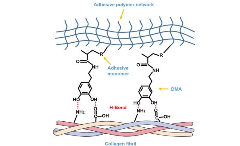
The possible rationale of DMA as a functional monomer in dentin bonding. DMA could be deemed as a “bridge” connecting the upper adhesive network via carbon-carbon double bond polymerization and lower dentin collagen fibrils via hydrogen bond, unifying them as one whole structure. Credit: The University of Hong Kong
Leading researchers from the Faculty of Dentistry, the University of Hong Kong (HKU), Wuhan University (WHU), and the Peking University Shenzhen Hospital have found that a compound found in mussels helps increase the durability of a dental filling.
In a journal article published in Materials Today Bio titled "Enhancing resin-dentin bond durability using a novel mussel-inspired monomer," they explain why this is a promising clinical finding for the future of dental filling treatments.
A dental filling is commonly used to restore tooth decay and broken teeth. Its durability highly depends on the longevity and stability of the bond between the compound (resin) and the hard tissue of the tooth (dentin). Here is where mussels play a role.
Small shellfish widespread in the marine environment, mussels offer unique wet adhesion properties which have long been of interest to the scientific community. Thus, the interaction between mussel plaques and substrates under humid environments has been extensively studied for insights on potential clinical applications. The study revealed that a compound found in an adhesive protein in mussels could strengthen the resin-dentin bond.
"Mussels need to maintain their adhesiveness under harsh marine environments, including humidity, drastic change of water temperature and pH value, sudden shocks and so on. These are similar to the daily activities that happen in the oral cavity. Our research aimed to understand the adhesive properties of the compounds from mussels, which may improve the durability and longevity of dental fillings," explained Professor Cynthia Kar Yung Yiu, clinical professor in pediatric dentistry, HKU who is leading the research team.
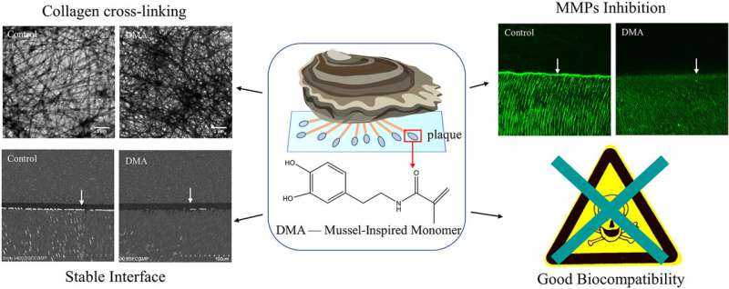
Comparison of the resin-dentin interface using DMA added adhesive and conventional dental adhesive. (set of photos on the left) DMA added adhesive results in a more stable interface. (set of photos on the right) In nanoleakage evaluation, in the control group, silver particles were observed spreading along the resin-dentin interface and infiltrating inside dentinal tubules after aging.
In a usual dental filling procedure, the dentist first removes the decayed tooth structure and fills the cavity with a tooth-colored restoration using a dental adhesive to glue the filling to the tooth structure. However, the durability of this bond can be affected by several factors, such as the humidity inside the oral cavity and repeated mechanical stress induced by chewing. Therefore, it remains a clinically significant challenge for the dentist as well as the patient as it leads to frequent replacement of the dental fillings at extra costs.
The study revealed that the wet adhesive property of mussels is attributed to the amino acid Dopa which they secrete. Based on the result, the team successfully applied N- (3,4-dihydroxyphenethy) methacrylamide (DMA), a mussel-derived compound, as a dental adhesive. The team further tested the durability of this resin-dentin interface versus the new DMA bond.
The control group and those with distinct concentrations of DMA underwent different tests including thermocycling aging, a process where dental materials are exposed to varied temperatures. The international standard for testing dental adhesives requires test specimens to be held repeatedly first in 5 °C cold water and then in 55 °C hot water for a large number of cycles. The results after subsequent testing invariably show a decrease in adhesive strength.
The researchers then employed the nanoleakage evaluation method in which an acid is added to measure the quality of the bond. The team used a silver nitrate solution to observe the patterns of nanoleakage.
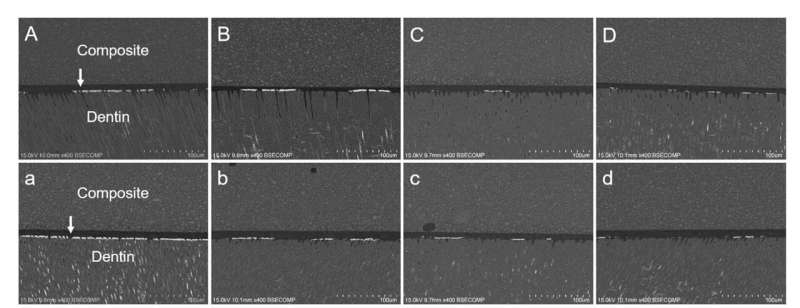
Images of nanoleakage expression from different groups. (A–D) The immediate groups. (a–d) The 10,000 times thermocycling aged groups. The first column is the Control. Groups on the right are treated with increasing concentrations of DMA. In the control group, nanoleakage deposition increased from 36.57% to 50.41% after thermoaging, while no obvious change could be detected for the DMA-treated groups. Credit: The University of Hong Kong
In the resin-dentin interface, the thermocycling aging process caused the formation of cracks and fissures which further led silver particles to infiltrate and settle along the bonded interface. The silver deposition therefore clearly reflected the water-filled and destructed areas along the interface. In the control group, silver particles were observed spreading along the resin-dentin interface and infiltrating inside dentinal tubules after aging (nanoleakage deposition increased from 36.57% to 50.41%) . On the contrary, no obvious change could be detected for the DMA-treated groups (nanoleakage deposition around 20%). The team thus deduced that DMA could strengthen the resin-dentin bond and its durability and is believed to increase the longevity of a dental filling.
"This research discovered that DMA is effective in strengthening the resin-dentin bond and improves its durability. The cytotoxicity is also similar to the resin monomers in traditional dental adhesives. It is believed that this compound may be commercialized in the future," said Dr. Tsoi.
More information: Kang Li et al, Enhancing resin-dentin bond durability using a novel mussel-inspired monomer, Materials Today Bio (2021). DOI: 10.1016/j.mtbio.2021.100174
Provided by The University of Hong Kong


