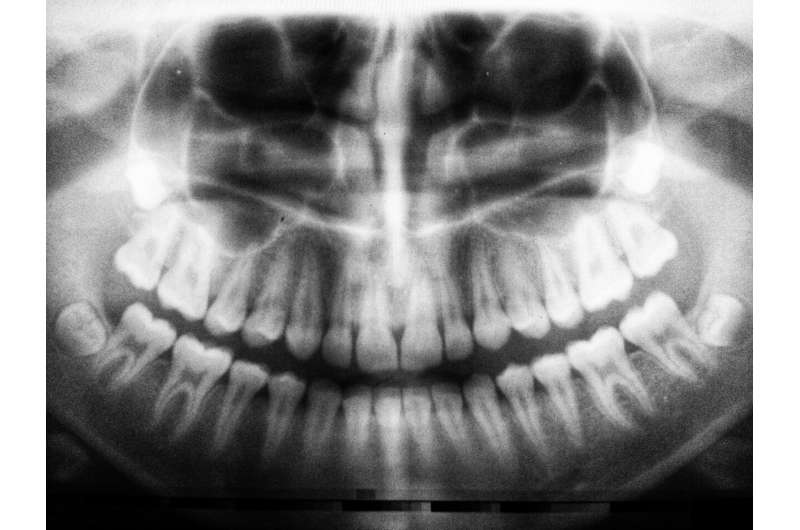
Artificial intelligence shows promise for interpreting dental X-rays

A deep learning algorithm successfully detects periodontal disease from 2D bitewing radiographs, according to research presented at EuroPerio10, the world's leading congress in periodontology and implant dentistry organized by the European Federation of Periodontology (EFP).
"Our study shows the potential for artificial intelligence (AI) to automatically identify periodontal pathologies that might otherwise be missed," said study author Dr. Burak Yavuz of Eskisehir Osmangazi University, Turkey. "This could reduce radiation exposure by avoiding repeat assessments, prevent the silent progression of periodontal disease, and enable earlier treatment."
Previous studies have examined the use of AI to detect caries, root fractures and apical lesions but there is limited research in the field of periodontology. This study evaluated the ability of deep learning, a type of AI, to determine periodontal status in bitewing radiographs.
The study used 434 bitewing radiographs from patients with periodontitis. Image processing was performed with u-net architecture, a convolutional neural network used to quickly and precisely segment images. An experienced specialist physician also evaluated the images using the segmentation method. Assessments included total alveolar bone loss around the lower and upper teeth, horizontal bone loss, vertical bone loss, furcation defects, and calculus around maxillary and mandibular teeth.
The neural network identified 859 cases of alveolar bone loss, 2,215 cases of horizontal bone loss, 340 cases of vertical bone loss, 108 furcation defects, and 508 cases of dental calculus. The success of the algorithm at identifying defects was compared against the physician's assessment and reported as sensitivity, precision and F1 score, which is the weighted average of sensitivity and precision. For sensitivity, precision and F1 score, 1 is the best value and 0 is the worst.
The sensitivity, precision and F1 score results for total alveolar bone loss were 1, 0.94 and 0.96, respectively. The corresponding values for horizontal bone loss were 1, 0.92 and 0.95, respectively, while AI could not identify vertical bone loss. For dental calculus, the sensitivity, precision and F1 score results were 1.0, 0.7 and 0.82, respectively, and for furcation defects the corresponding values were 0.62, 0.71 and 0.66, respectively.
Dr. Yavuz said: "Our study illustrates that AI is able to pick up many types of defects from 2D images which could aid in the diagnosis of periodontitis. More comprehensive studies are required on larger data sets to increase the success of the models and extend their use to 3D radiographs."
He concluded: "This study provides a glimpse into the future of dentistry, where AI automatically evaluates images and assists dental professionals to diagnose and treat disease earlier."


