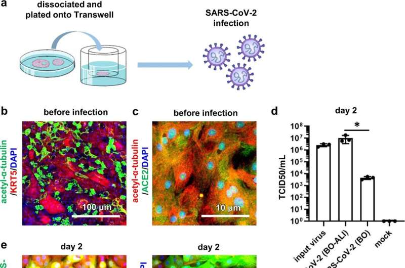
SARS-CoV-2 infection experiments in BO-ALI. a BO-ALI were infected with SARS-CoV-2 (1.3 × 10⁵ TCID50/well) and then cultured with differentiation medium for 2 days. b Immunofluorescence analysis of KRT5 (red) and acetylated α-tubulin (green) in uninfected BO-ALI. Nuclei were counterstained with DAPI (blue). c Immunofluorescence analysis of ACE2 (green) and acetylated α-tubulin (red) in uninfected BO-ALI. Nuclei were counterstained with DAPI (blue). d The amount of infectious virus in the supernatant of infected BO or BO-ALI was measured by the TCID50 assay. Statistical significance was evaluated using one-way ANOVA followed by Tukey’s post hoc test (*P < 0.05). Data represent the mean ± SD from three independent experiments. e Immunofluorescence analysis of SARS-CoV-2 Spike protein (SP) (green) and acetylated α-tubulin (red) in uninfected BO-ALI 2 days after the infection. Immunofluorescence analysis of SARS-CoV-2 SP (green) and KRT5 (red) in uninfected BO-ALI. Nuclei were counterstained with DAPI (blue) 2 days after the infection. f Immunofluorescence analysis of SARS-CoV-2 SP (green) and acetylated α-tubulin (red) in infected BO-ALI 7 days after the infection. Immunofluorescence analysis of SARS-CoV-2 SP (green) and KRT5 (red) in infected BO-ALI 7 days after the infection. Nuclei were counterstained with DAPI (blue). g Immunofluorescence analysis of KRT5 (red) and acetylated α-tubulin (green) in BO-ALI 15 days after the infection. Nuclei were counterstained with DAPI (blue). Panels b, c, e–g are representative of three independent experiments. Credit: Communications Biology (2022). DOI: 10.1038/s42003-022-03499-2
The group led by Dr. Kazuo Takayama developed two in vitro models to study SARS-CoV-2, a bronchial organoids model (BO) and a BO-derived air-liquid interface model (BO-ALI), and showed that they can be used for drug screening for infectious diseases including COVID-19.
An in vitro lung model that faithfully reproduces the human respiratory organ is essential for conducting COVID-19 drug screening. BO and alveolar organoids are excellent tools for faithfully mimicking the human respiratory organ and are expected to be applied to COVID-19 research.
In this study, the research team developed two human bronchial models, BO and BO-ALI. SARS-CoV-2 infection experiments confirmed that the infection efficiency in BO-ALI was more than 2,000 times higher than that in BO. The researchers confirmed the anti-viral effect of therapeutic agents such as remdesivir, molnupiravir, and camostat, using BO-ALI. Comparative analyses of eight SARS-CoV-2 variants, including the omicron strain, showed that the infection efficiency in BO-ALI of the omicron was slightly lower than those of the other variants.
Next, in attempting to identify the cells that SARS-CoV-2 infects, the researchers found that the virus efficiently infects ciliated cells but rarely infects basal cells. They also discovered that nearly all ciliated cells died subsequently whereas basal cells survived and differentiated into bronchial epithelial cells, including ciliated cells. The regenerative capacity of basal cells was dependent on fibroblast growth factor 10 (FGF10).
These findings suggest that BO-ALI produced from BO can be used to evaluate COVID-19 therapeutics, analyze SARS-CoV-2 variants, and study bronchial tissue regeneration.
The results of this study were published online in Communications Biology on May 30, 2022.
More information: Emi Sano et al, Cell response analysis in SARS-CoV-2 infected bronchial organoids, Communications Biology (2022). DOI: 10.1038/s42003-022-03499-2
Journal information: Communications Biology
Provided by Kyoto University


