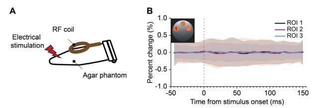
Sham DIANA experiment. (A) Illustration of a sham DIANA experiment with electrical stimulation using an agar phantom instead of an in vivo mouse on a 9.4 T scanner. (B) Time series of percent signal changes in DIANA responses extracted from three regions-of-interest (ROI, defined in the subfigure) in a 1 mm coronal slice. Data from 400 trials were averaged, where the number of trials was determined by considering the group-averaged results, e.g., 40 trials/mouse × 10 mice in Fig. 2D, for comparison of temporal fluctuation. The red vertical dotted line indicates the electrical stimulation onset time. Mean values were displayed as solid lines and standard deviations in shades (black, ROI 1; magenta, ROI 2; blue, ROI 3). Credit: Science (2022). DOI: 10.1126/science.abh4340
A team of researchers affiliated with multiple institutions in the Republic of Korea has developed a new way to use magnetic resonance imaging (MRI) for noninvasive tracking of the propagation of brain signals on millisecond timescales. In their paper published in the journal Science, the group describes how they developed the new technology, its features and how well it worked when tested on mice. Timo van Kerkoerle and Martijn Cloos with Université Paris-Saclay and the University of Queensland, respectively, have published a Perspectives piece in the same journal issue outlining the work done by the team on this new effort.
MRI works by sending a magnetic field along with radio waves through tissue to produce images of the tissue under study. It works because different types of tissue have different properties. MRI is also used in different ways depending on the part of the body under study. Blood-oxygen-level-dependent functional MRI is used to obtain images of the brain of a living person—the technique allows for observing changes in blood flow in the brain that serve as a stand-in for an increase in neuronal activity. In practice, blood-oxygen-level-dependent functional MRI produces multiple images over time, generally over several seconds. In this new effort, the researchers have modified the way an MRI brain scan is performed.
The new technique, called direct imaging of neuronal activity (DIANA) works by modifying a traditional MRI machine to create images faster—on the millisecond scale—potentially at the speed of thought. It does so by creating partial images that are combined afterward to create a high-resolution image that represents an instance of brain activity. After a single scan, multiple high-resolution images are created that show the state of the brain as it responds to a stimulus, such as a jolt of electricity applied to the whiskers of test mice. By generating images so quickly, the technique allows researchers to observe the propagation of brain signals from one part of the brain to another in a noninvasive manner.
The new technique has thus far only been tested in mice, but the researchers are already calling it a game-changer, suggesting it could transform the way that scientists study the brain—and could lead to a new understanding of how it works.
More information: Phan Tan Toi et al, In vivo direct imaging of neuronal activity at high temporospatial resolution, Science (2022). DOI: 10.1126/science.abh4340
Timo van Kerkoerle et al, Creating a window into the mind, Science (2022). DOI: 10.1126/science.ade4938
Journal information: Science
© 2022 Science X Network


