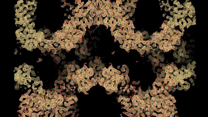
Using cryo-electron microscopy, researchers obtained a detailed map (shown in orange) of the chain-like tankyrase structure. By interpreting this map and reconstructing the tankyrase molecules amino acid by amino acid (red), they deciphered how tankyrase is activated by ‘self-assembling’. Credit: Sebastian Guettler, ICR.
Scientists have revealed the inner workings of a key protein involved in a wide range of cellular processes—potentially paving the way for better and less toxic cancer drugs.
Using Nobel Prize-winning microscopy techniques, the researchers revealed how the tankyrase protein switches itself on and off by self-assembling into 3D chain-like structures.
Their study, published in the journal Nature, reveals crucial structural insights into the elusive but important tankyrase protein, which plays a particularly important role in helping drive bowel cancer.
Scientists at The Institute of Cancer Research, London, believe their research will open the door to new types of cancer treatment that can control tankyrase more precisely than is currently possible, with fewer side effects.
The fundamental discovery could have implications for treating various cancers, as well as diabetes and inflammatory, cardiac and neurodegenerative diseases.
Tankyrase is an important protein that supports 'Wnt signaling'—signals that are essential for the body to maintain stem cells and carry out processes like cell division and development but, when uncontrolled, can fuel bowel cancer, among others. Tankyrase also controls other cell functions critical to cancer, such as the maintenance of the ends of chromosomes, the telomeres.
Unlike the PARP1 protein from the same 'PARP family', tankyrase remains poorly understood. While drugs blocking PARP1 have already made it to the clinic, scientists still don't fully comprehend how tankyrase is switched on, how it functions or how to block it without leading to unwanted side effects.
In this study, scientists draw parallels between the activation mechanism of PARP1 and tankyrase for the first time. They suggest that, similarly to PARP1, tankyrase works by being recruited to a specific site and 'self-assembling', clustering and changing its 3D structure to activate itself and perform its function.
In the last decade, scientists have developed drugs to block tankyrase in an attempt to treat bowel cancer—but because Wnt signaling is involved in a wide range of processes, the drugs led to too many side effects for them to reach clinical trials.
To really understand how tankyrase inhibitors work and how to develop less toxic treatments, scientists at the ICR set out to discover new structural information using cutting-edge cryo-electron microscopy. This extremely powerful type of microscopy freezes samples at -180°C to enable minute details of protein shape to be imaged.
The approach allowed them to visualize and capture how tankyrase 'self-assembles' into fibers—chain-like structures—and why fiber formation is needed for tankyrase to activate itself.
Researchers believe the 'domains'—specific regions of the protein associating with different functions—that allow tankyrase to assemble and disassemble into different structures are exciting targets for future cancer drugs. They also believe that, depending on which structural domains drugs bind to, not all tankyrase inhibitors will affect Wnt signaling in the same way.
The hope is that researchers will be able to design structurally different tankyrase inhibitors—ones that are safer and more effective, which are urgently needed for treating bowel cancer and other diseases with which tankyrase has been linked.
Study leader Professor Sebastian Guettler, Deputy Head of the Division of Structural Biology at The Institute of Cancer Research, London, said, "Our study has provided vital new information about a particular protein molecule called tankyrase, which plays an important role in bowel cancer and other diseases but has so far eluded our understanding. We're playing catch up—we have all these drugs to block tankyrase being created, but we don't have enough basic understanding to use them as treatments."
"We have shown how tankyrase is switched on and can go from a 'lazy' enzyme to an active one. If we can create better, less toxic drugs to control this process, we could pave the way for an effective bowel cancer treatment in the future."
Professor Kristian Helin, Chief Executive of The Institute of Cancer Research, London, said, "These fundamental findings help us understand how the extremely important tankyrase protein works within cells. Almost all bowel cancers have hyperactive Wnt-signaling that operates through tankyrase, and they could therefore potentially be treated with drugs targeting it."
"I am hopeful these key advances in our understanding of tankyrase will help us overcome the limitations of currently available drug candidates—hopefully bringing us a step closer to a new targeted bowel cancer treatment. Tankyrase is also responsible for regulating a wide range of processes relevant to a variety of diseases, not just cancer, so this research could have broad implications."
Dr. Marianne Baker, Research Information Manager at Cancer Research UK, said, "PARPs can help cancer cells fix their damaged DNA, so they're crucial targets for cancer-killing drugs. We're proud to have supported this research that builds knowledge of the less studied tankyrase PARPs and could help pave the way for new treatments in the future."
"This paper is an example of important discovery research that deepens our understanding of biology, which is vital for designing new cancer drugs."
"It also builds on Cancer Research UK's successful history with PARP inhibitors. In the 1990s, Cancer Research UK-funded scientists at the ICR played a key role in developing drugs that inhibit PARP proteins and stop cancer cells repairing themselves. Tens of thousands of people across the world now receive these treatments."
More information: Sebastian Guettler, Structural basis of tankyrase activation by polymerisation, Nature (2022). DOI: 10.1038/s41586-022-05449-8
Journal information: Nature
Provided by Institute of Cancer Research


