This article has been reviewed according to Science X's editorial process and policies. Editors have highlighted the following attributes while ensuring the content's credibility:
fact-checked
peer-reviewed publication
trusted source
proofread
COVID-19 associated with fetal brain hemorrhages

New research from the Institute of Psychiatry, Psychology & Neuroscience (IoPPN) at King's College London has found evidence of small hemorrhages in the brain tissue of fetuses during the peak of COVID-19 cases in the UK.
The research, published in Brain, found that the hemorrhages are linked to a reduction in blood vessel integrity. The cause of these hemorrhages is unclear, however possible explanations might be as a direct consequence of the infection or an indirect consequence of the maternal immune response. The study suggests that COVID-19 might affect the fetal brain during the earliest stages of gestation, highlighting a need for further study into the potential impact on subsequent neurological development.
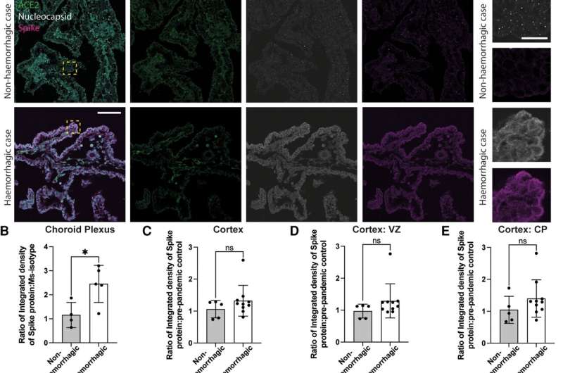
The researchers studied 26 samples of human fetal tissue with observed hemorrhages from a total of 661 samples collected between July 2020 and April 2022. It was established that the COVID-19 virus was present in all of the hemorrhagic samples.
The majority of the hemorrhagic samples came from donated fetal tissue between the late first and early second trimester of gestation—a particularly important period of human fetal brain development during which the tight junctions between endothelial cells of the blood vessels increase to form the blood brain barrier, the semipermeable barrier that protects the brain from foreign substances.
Upon further study, the integrity of the blood vessels within the hemorrhagic samples was found to be considerably lower than the non-infected samples, ultimately providing an explanation as to why bleeds could be seen in the samples.
"While hemorrhages do occasionally occur in developing brains, it is extremely unusual for there to be this many instances within a 21 month period. It is now of the utmost importance that we follow up with children that were prenatally exposed to COVID-19 so that we can establish if there are any long-lasting neurodevelopmental effects," said Dr. Katie Long, the study's lead investigator from King's IoPPN.
Marco Massimo, the study's first author from King's IoPPN said, "Our findings suggest that there is an association between the early development of human fetal brain tissue and vulnerability to infection from COVID-19."
Professor Lucilla Poston CBE, Professor of Maternal & Fetal Health at King's College London, said, "We know that severe viral infection may influence the fetal brain, but this important study is the first to suggest that this may occur in pregnancies affected by COVID infection. Whatever the cause, a direct effect of the virus or an indirect consequence of maternal infection, this study highlights the need for pregnant women to be vaccinated against COVID-19, thus avoiding complications for both mother and baby."
Dr. Long concludes, "While we haven't yet been able to establish clear causation, we certainly feel that it highlights a need for expectant mothers to take extra precautions at a time when cases are on the rise."
More information: Marco Massimo et al, Haemorrhage of human foetal cortex associated with SARS-CoV-2 infection, Brain (2023). DOI: 10.1093/brain/awac372


