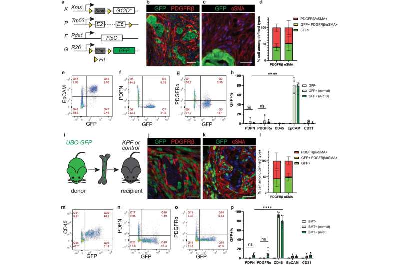
Epithelium and bone marrow minimally contribute to pancreatic cancer associated fibroblasts. a Schematic of the KPFG mouse model. b–d Co-immunostaining in the KPFG pancreas and quantification of cells that are either single positive or double positive for defined markers. n = 7 mice for PDGFRβ analysis; n = 8 mice for αSMA analysis. e–h Flow cytometry analysis of dissociated KPFG pancreas and quantification of GFP + cells within populations defined by markers. n = 4 mice for GFP-; n = 3 mice for GFP + (normal); n = 9 mice for GFP + (KPFG). ****p < 0.0001; ns for PDPN: p = 0.996; ns for PDGFRα: p = 0.923. i Schematic of the bone marrow transplant (BMT) experiment. j–l Co-immunostaining in KPF pancreas transplanted with GFP + bone marrow and quantification of cells that are either single positive or double positive for defined markers. n = 5 mice for PDGFRβ analysis; n = 3 mice for αSMA analysis. m–p Flow cytometry analysis of pancreas from KPF mice transplanted with GFP + bone marrow and quantification of GFP + cells within populations defined by markers. n = 4 non-transplanted mice; n = 5 transplanted normal mice; n = 6 transplanted KPF mice. Data are mean ± SD. ****p < 0.0001; ns for PDPN: p = 0.744; ns for PDGFRα: p = 0.340. h, p Two-way ANOVA test with Tukey’s multiple comparisons was performed. ns, not significant. Scale bars: 30 μm. Credit: Nature Communications (2023). DOI: 10.1038/s41467-022-34464-6
A type of cell that plays a key role in pancreatic cancer can trace its origin back to a structure that forms during embryonic development, new research at MUSC Hollings Cancer Center shows. This new data, published recently in Nature Communications, is the first to show the cellular origin of normal pancreatic fibroblasts and cancer-associated fibroblasts (CAFs) that influence tumor progression. The research findings open the door for new therapeutic targets for pancreatic cancer, which remains challenging to treat.
Pancreatic ductal adenocarcinoma, which accounts for more than 90% of pancreatic cancers, is one of the deadliest types of cancer. Despite accounting for only about 3% of all cancers in the U.S., it causes about 7% of cancer deaths. According to the American Cancer Society, more than 50,000 people are expected to die from pancreatic cancer in the U.S. in 2023.
"More effective therapeutics are urgently needed for patients with pancreatic cancer," said Lu Han, Ph.D., lead author of the publication and a postdoctoral fellow in the laboratory of Hollings researcher Michael Ostrowski, Ph.D., the WH Folk Endowed Professor in Experimental Oncology. "Most patients die within five years of their diagnosis. We aimed to learn the basic biology of pancreatic cancer so we can provide information to lead to more effective treatments."
Ostrowski's research focuses on understanding the molecular interactions in breast and pancreatic cancer to make fundamental scientific discoveries that lead to future improvements in patient outcomes. "We are still learning about the cells that make up the tumor microenvironment. New therapies often take decades to develop because we first must understand tumor biology. Our recent discovery about the origin of cancer-associated fibroblasts will help the pancreatic cancer field to progress," Ostrowski said.
While pancreatic cancer is driven by tumor cells, fibroblasts are cells that control the microenvironment around the tumor. "A healthy pancreas contains very few fibroblasts. However, there is a drastic increase in the number of fibroblasts when a tumor forms in the pancreas and fibroblasts play important and complex roles in how the disease progresses," Han said.
Targeting CAFs holds exciting potential for therapeutic benefits. Han said that knowing the basic biology of this cell type is the initial step. Many studies show that there may be multiple types of CAFs, which may play different roles in either promoting or prohibiting cancer progression. "Understanding the specific factors within each CAF subtype will help us direct future therapeutic design. Ideally, we want to be able to promote the CAFs that reduce cancer cell growth and inhibit the tumor-promoting CAFs," said Han.
They first focused on determining the origin of these cells. "Other researchers have proposed that the different CAF subtypes found in pancreatic cancers come from different origins, meaning that they develop from different cells," Han said. The potential CAF origins include pancreatic epithelial cells, the bone marrow and pancreatic resident fibroblasts—fibroblasts in the normal adult pancreas. They formally tested that hypothesis by engineering elegant mouse genetic models of pancreatic cancer.
"What we found was a bit surprising. The bone marrow and epithelial cells only had minimal contributions to CAFs. We found that both resident fibroblasts and CAFs came from the splanchnic mesenchyme, a layer of cells in an embryo during early development," Han said. This new finding about the fetal origin of the pancreatic fibroblasts suggested that the different CAF subtypes found in pancreatic cancer are likely caused by the tumor microenvironment driving the diversification of the fibroblasts.
Next, Han studied whether the CAFs also had the same gene signature of their fetal origin. Figuring out the gene signature similarities could offer novel insights into CAF heterogeneity and the complex roles CAFs play in pancreatic cancer progression.
The gene signature studies revealed two distinct groups of genes. The normal pancreatic fibroblasts shared a group of genes with the splanchnic mesenchyme that is potentially important for normal organ maintenance. The CAFs shared a different group of genes, which potentially play a role in promoting rapid cell multiplication. This observation was seen in their mouse models and human pancreatic tissues, suggesting that some fetal development programs are turned back on in the fibroblast lineage during tumor development.
"During tumor development, the splanchnic-derived fibroblasts quickly multiply and become a large portion of the pancreatic cancer tissue mass. The importance of the fetal development programs in the CAFs is a really exciting direction and has not been explored before this study," said Han, who wants to continue pushing the boundaries of cancer research once she becomes an independent researcher.
The results give a new direction for pancreatic cancer researchers by providing valuable insights into fibroblast biology during pancreatic cancer progression. Han and Ostrowski's laboratory are diving into more studies to understand the functional importance of the fetal development program in CAFs, which will be translated into better-targeted therapeutics in the future.
More information: Lu Han et al, The splanchnic mesenchyme is the tissue of origin for pancreatic fibroblasts during homeostasis and tumorigenesis, Nature Communications (2023). DOI: 10.1038/s41467-022-34464-6
Journal information: Nature Communications
Provided by Medical University of South Carolina


